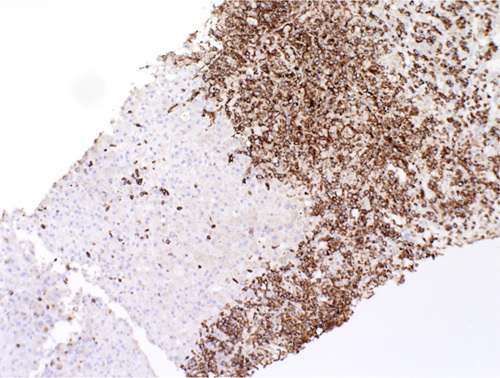
FIGURE 2.

CD3 immunohistochemical staining of the atypical cells within the biopsy. These cells show strong diffuse membranous expression of CD3 consistent with T-cell origin. There is loss of other pan T-cell antigens (CD5 and CD7) and “display a double-negative” phenotype (CD4 and CD8 negative). The lack of clear immunophenotypic pattern classifies this as peripheral T-cell lymphoma not otherwise specified.
