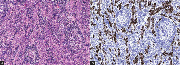
Figure 2.
(a) Photomicrograph of the resected mass shows well-formed ducts lined by cytologically bland cuboidal epithelial cells and accompanying lymphoid infiltrate forming reactive follicles with germinal centres (H&E, ×100). (b) Photomicrograph of the ductal epithelium shows diffuse positivity for CK19 immunohistochemical stain (×100).

