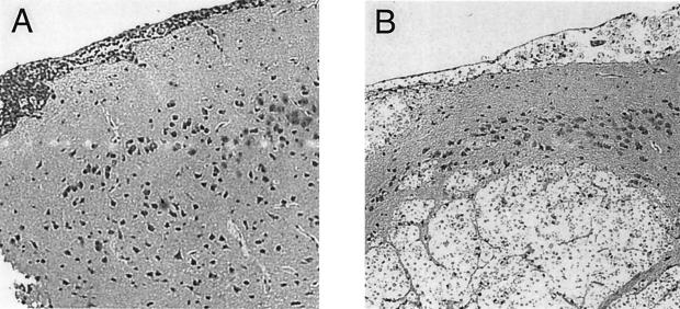
FIG. 9.
Histology of brain sections 10 weeks post-intratracheal infection with C. neoformans 145A. Magnification, ×33. (A) WT mice. Note the well-organized, local inflammatory cell infiltrate colocalized with cryptococci and the good separation of infected areas of meninges from uninfected brain tissue. (B) MIP-1α KO mice. Note the swelling and infiltration of the meninges and multiple intracerebral lesions, the round foci expanding within the brain with numerous cryptococci with large capsules, no or few inflammatory cells.

