FIGURE 3.

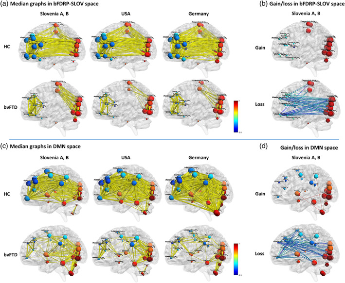
Functional connectivity within (a) bFDRP‐SLOV and (c) default mode network (DMN) vector space in all the three populations from Slovenia, USA, and Germany. The right panels (b and d) represent gain and loss of connections in bvFTD patient compared to HC in patients from Slovenia. Functional connectivity is presented for normal controls (NC) and behavioral variant frontotemporal dementia (bvFTD) patients. Reorganization of network is seen in both network spaces; however, in bFDRP‐SLOV space (a) the network is completely disconnected while in the DMN space (c) frontal areas remain connected with other parts of the network through middle/posterior cingulate, precentral, and supramarginal nodes. Edge thickness corresponds to the magnitude of the correlation. All the edges exceeding |r| > .6 are presented. (b, d) Analysis of gain and loss of connection between HC and bvFTD in bFDRP‐SLOV and DMN vector spaces presented on data from cohorts Slovenia A and B. (Nodes are color‐coded according to the CJDRP weights; the diameter of nodes corresponds to eigenvector centrality [see Section 2].)
