
FIGURE 1.
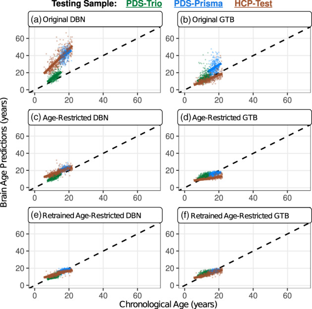
Brain age predictions were consistently overestimated, especially for scans acquired from Siemens Prisma scanners. This can be observed by how different the colored regression lines are relative to the black dashed line, which represents a perfect model fit (coefficient = 1; y‐intercept = 0). These biases were most pronounced when applying the original model variations (a & b). Attempts to restrict the age range of the training sample (c & d) and subsequently retrain each model (e & f) had varying levels of improvements, but issues of prediction bias persisted (Table 2). The PDS‐trio sample is represented by green circles, the PDS‐Prisma by blue triangles, and the HCP‐test by brown squares