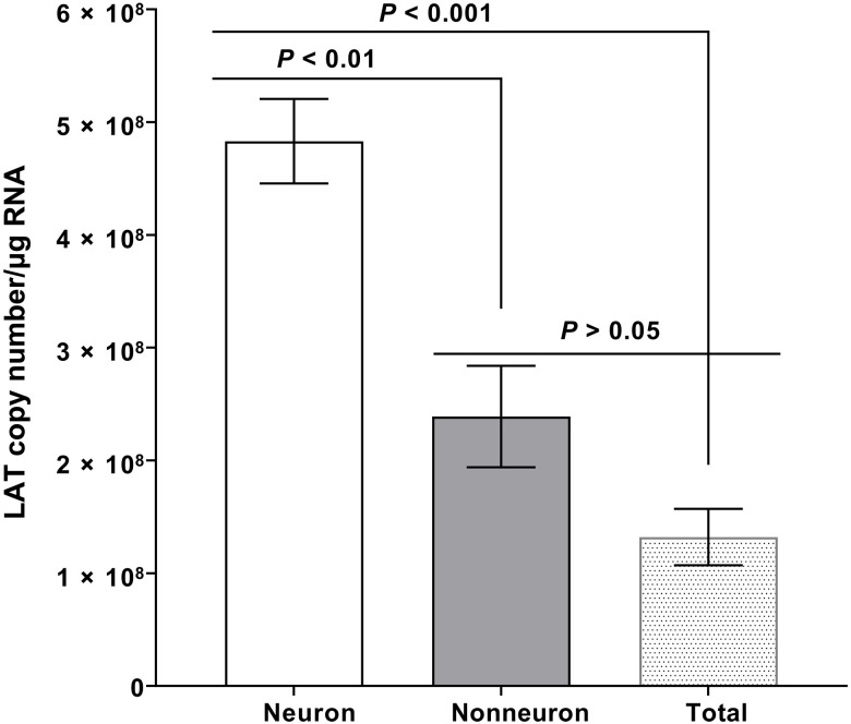
Fig. 1. Detection of LAT in the nonneuronal fraction of latently infected TG.
Wild-type (WT) mice were ocularly infected with 2 × 10⁵ PFU per eye of HSV-1 strain McKrae without corneal scarification. TG were harvested on day 35 PI. Four latently infected TG from two mice were pooled together and dissociated into a single-cell suspension by digesting with collagenase D. Neuronal and nonneuronal fractions were isolated using the Miltenyi Biotec neuron isolation kit, as described in the company protocol. Briefly, dissociated cells were stained with the antibody cocktail, mixed with magnetic beads, and neuronal and nonneuronal cells were collected. Total RNA was extracted from each fraction, and unfractionated RNA from individual TG was used as a control. LAT copy number was measured by qRT-PCR using a standard curve generated from pGEM5317, as we described previously (73). Glyceraldehyde-3-phosphate dehydrogenase (GAPDH) expression was used to normalize relative levels of LAT RNA expression. Each bar represents the mean LAT copy number/μg RNA ± SEM from 12 mice for nonneuronal RNA and neuronal RNA and from 12 mice for total TG RNA.

