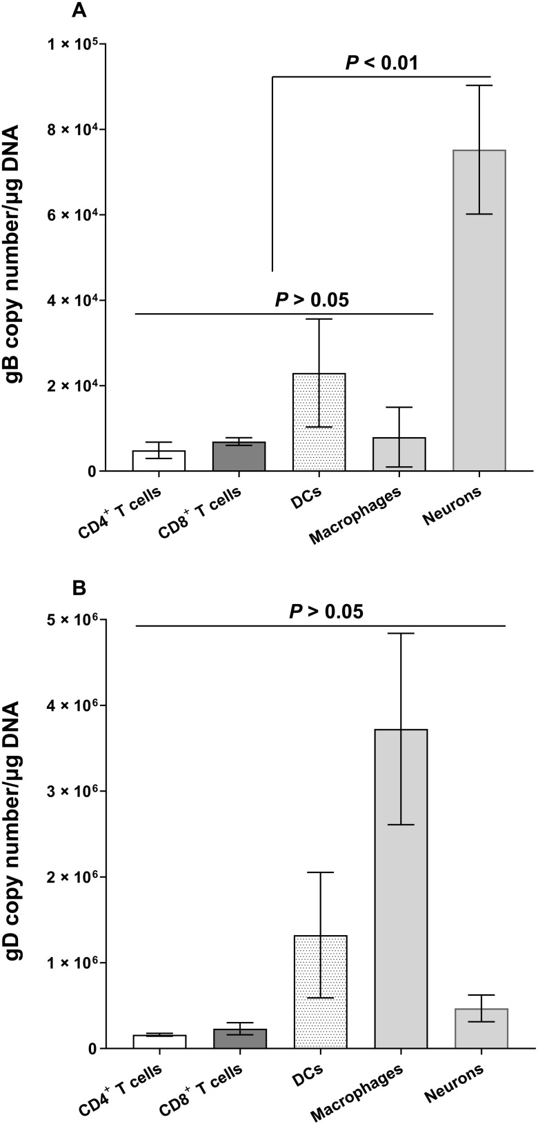
Fig. 6. Detection of gB and gD DNA in neuronal and nonneuronal fractions of latently infected TG.
WT mice were infected ocularly with HSV-1 McKrae as described in Fig. 1 legend. TG were isolated on day 35 PI, and eight TG from four mice were pooled and dissociated to single cells. Immune cells enriched by Percoll density gradient were stained with CD45, CD3, CD4, CD8, F4/80, and CD11c antibodies. CD4, CD8, F4/80, and CD11c populations were sorted, and genomic DNA from each population was purified. gB (A) and gD (B) copy numbers were measured by qPCR, and GAPDH was used as internal control. Experiments were repeated three times.

