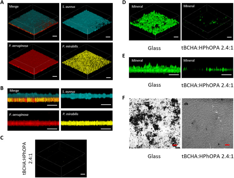
Fig. 5. Resistance of the tBCHA:HPhOPA 2.4:1 copolymer to mixed-species biofilm formation and biomineralization in AU.
A polymicrobial biofilm was allowed to develop on glass or on the copolymer where colonization over the first 24 hours was with gfp-tagged Proteus, followed by mCherry-tagged Ps. aeruginosa PAO1 (red) and cfp-tagged S. aureus SH1000 (blue) in a 1:1 ratio. After a further 48-hour incubation, the samples were observed via confocal microscopy. (A) Three-dimensional (3D) representation of the mixed-species biofilm on glass. (B) Transverse view of the mixed-species biofilm. (C) 3D representation showing (C) the lack of a mature biofilm on the copolymer. (D) 3D representation and (E) transverse view of biomineralization on glass and copolymer. (F) Bright-field images of biomineralization on glass and copolymer. Scale bars, 50 μm.

