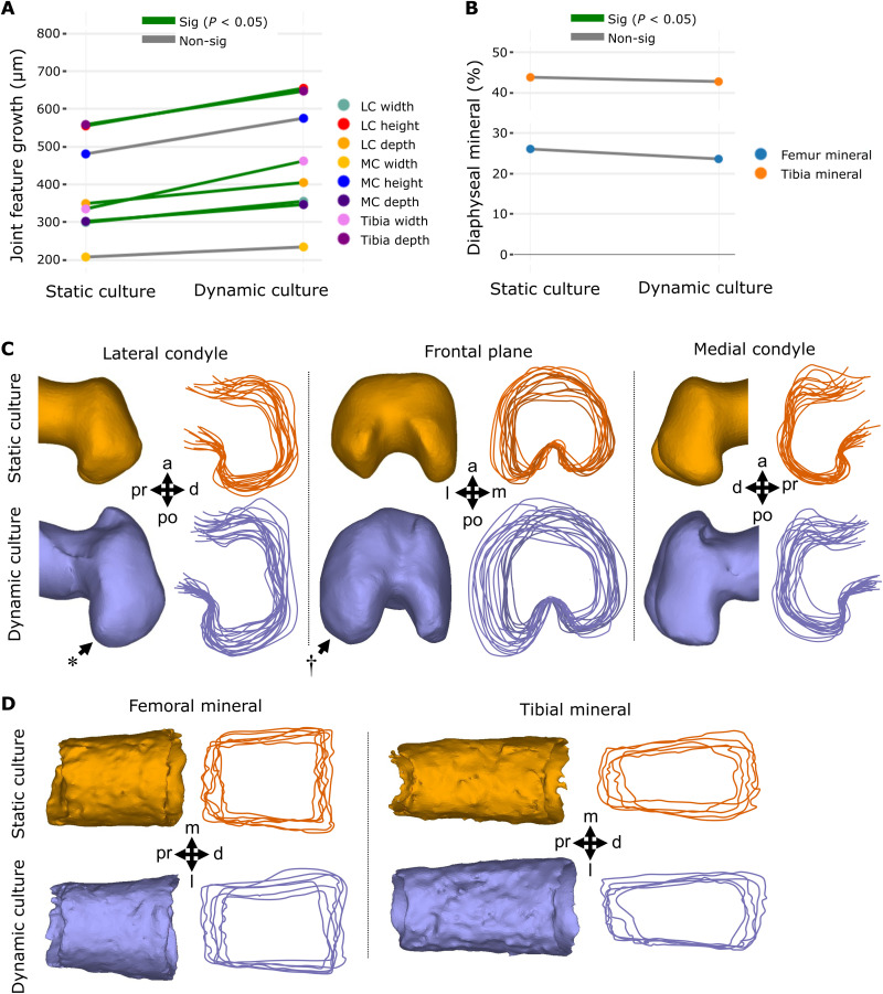
Fig. 2. Dynamic loading of cultured mouse embryo hindlimbs promotes joint cartilage feature growth and shape but not the extent of mineralization or bone collar shape.
(A) Average paired differences in growth for joint cartilage features (each line represents one of eight features) in static versus dynamic culture (n = 8 limbs per group). Green lines, significant (P < 0.05) feature differences; gray lines, nonsignificant feature differences. (B) Average extents of femur (n = 6) and tibia mineralization (n = 6) in static and dynamic cultured paired limbs. (C) Representative samples and joint shape contours for distal femora cultured in static and dynamic conditions. Arrows indicate regions of increased growth and shape development. * represents more prominent posterior curl observed in dynamically loaded limbs. † represents larger lateral expansion found in the lateral condyle of dynamically loaded limbs. (D) Representative samples and outlines of femoral and tibial bone collars from limbs cultured in static and dynamic conditions. Pr, proximal; d, distal; a, anterior; po, posterior; m, medial; l, lateral; MC, medial condyle; LC, lateral condyle.

