. 2023 Jan 10;12:e85837. doi: 10.7554/eLife.85837

Figure 3. Disruption of palmitoylation of DNAJC5 inhibited α-syn secretion.

(A) Inhibition of DNAJC5 palmitoylation by 2-bromopalmitic acid (2-BA) or introduced mutation L115R. Cellular fractionation was performed with HEK293T cells transfected with WT DNAJC5 and treated with 10 μm 2-BA, or transfected with DNAJC5 L115R mutant. C, cytosol; M, membrane; PNS, post-nuclear supernatant; TFR, transferrin receptor. (B) Quantification of the percentage of P-DNAJC5 and NP-DNAJC5 in different conditions as shown in (A). Error bars represent standard deviations of three experiments. (C) α-syn secretion was blocked with 2-BA treatment. HEK293T cells transfected with indicated plasmids were treated with DMSO or 10 μm 2-BA. Media fractions were collected and secretion was evaluated by SDS-PAGE and immunoblot. (D) Palmitoylation of DNAJC5 was blocked in HEK293T cells treated with 2-BA. (E) Quantification of normalized α-syn secretion in HEK293T cells after 2-BA treatment. The quantification was based on immunoblot in (C) and (D). The α-syn secretion was calculated as the amount of α-syn in media divided by the amount in lysate. (F) Quantification of normalized DNAJC5 secretion in HEK293T cells after 2-BA treatment. The quantification was based on immunoblot in (C) and (D). The DNAJC5 secretion was calculated as the amount of DNAJC5 in media divided by the amount in lysate. (G) DNAJC5 L115R mutant reduced α-syn secretion compared with WT DNAJC5. Secretion assay with HEK293T cells transfected with indicated plasmids encoding DNAJC5 variant was performed similar to (C). (H) DNAJC5 L115R was non-palmitoylated in HEK293T cells. (I) Quantification of normalized α-syn secretion in HEK293T cells transfected with DNAJC5 L115R mutant. The quantification was based on immunoblot in (G) and (H). (J) Quantification of normalized DNAJC5 secretion in HEK293T cells transfected with DNAJC5 L115R mutant. The quantification was based on immunoblot in (G) and (H).

Figure 3—source data 1. Uncropped immunoblot corresponding to Figure 3.

Figure 3.

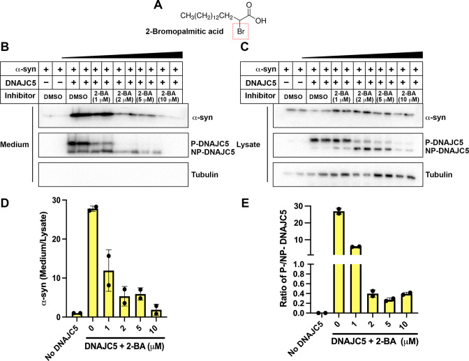
Figure 3—figure supplement 1. Dose-dependent inhibition of α-syn secretion by 2-bromopalmitic acid (2-BA).

Figure 3—figure supplement 1.

(A) Chemical structure of palmitoylation inhibitor 2-bromopalmitic acid (2-BA). The single bromo substituent at position 2 is highlighted by a red dashed square. (B) α-syn secretion in the medium was inhibited by 2-BA in a dose-dependent manner. About 10 μm 2-BA was serially diluted by DMSO into 5 μm, 2 μm, and 1 μm solution. HEK293T cells were first transfected with DNAJC5 and α-syn. After medium replacement, 2-BA of indicated concentration was added to cell culture. Media were collected after 36 hr and followed by sample preparation, SDS-PAGE and immunoblot. (C) With increasing concentration of 2-BA, P-DNAJC5 decreased and NP-DNAJC5 increased in HEK293T cells. (D) Quantification of normalized α-syn secretion upon increasing concentration of 2-BA. Quantification was based on immunoblot in (B) and (C). The α-syn secretion was calculated as the amount of α-syn in media divided by the amount in lysate. (E) Quantification of ratio of P-DNAJC5/NP-DNAJC5. The quantification was based on immunoblot in (C).
Figure 3—figure supplement 1—source data 1. Uncropped immunoblot corresponding to Figure 3—figure supplement 1.
Figure 3—figure supplement 2. Blockage of USP19-induced α-syn secretion by DNAJC5 L115R or L116Δ mutant.

Figure 3—figure supplement 2.

(A) USP19 induced α-syn secretion in the medium, which was further enhanced by WT DNAJC5 and blocked by two DNAJC5 palmitoylation-deficient mutants (L115R and L116Δ). (B) Both mutations, L115R and L115Δ, inhibited DNAJC5 palmitoylation in HEK293T cells. (C) Quantification of normalized α-syn secretion in HEK293T cell transfected with indicated constructs. The quantification was based on immunoblot in (A) and (B). α-Syn secretion was calculated as the amount of α-syn in media divided by the amount in lysate.
Figure 3—figure supplement 2—source data 1. Uncropped immunoblot corresponding to Figure 3—figure supplement 2.
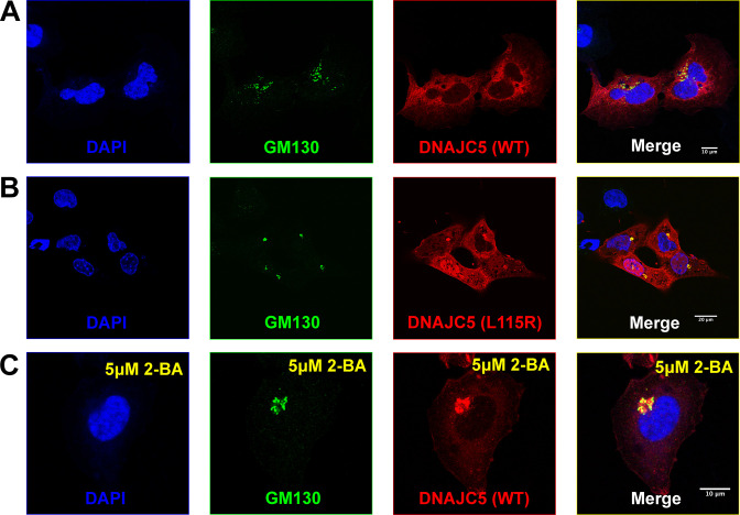
Figure 3—figure supplement 3. Golgi retention of DNAJC5 in secretion-deficient conditions.

Figure 3—figure supplement 3.

(A) Immunofluorescence (IF) images of U2OS cells transfected with wildtype (WT) DNAJC5-HaloTag. Before fixation, HaloTag TMR ligands were added to the cell culture to label DNAJC5. Golgi apparatus was visualized by IF using anti-GM130 antibody. Nuclei were stained with DAPI. Scale bar: 10 μm. (B) IF images of U2OS cells transfected with DNAJC5 (L115R)-HaloTag. Same staining procedure was performed as in (A). Scale bar: 20 μm. (C) IF images of U2OS cells transfected with WT DNAJC5-HaloTag and treated with 5 μM 2-BA. After transfection with WT DNAJC5-HaloTag, cells were incubated with 5 μM 2-BA for 1 day to block palmitoylation of DNAJC5. Same staining procedure was performed as in (A). Scale bar: 10 μm.