. 2023 Jan 10;12:e85837. doi: 10.7554/eLife.85837

Figure 5. Secretion of tandem α-syn oligomers and α-syn fused with thermostable helix-bundle protein.

(A) Schematic diagrams of tandem α-syn oligomers. α-syn protomers (yellow) were linked head to tail by flexible linker (green) to mimic increased size of α-syn oligomers. (B) Secretion of tandem α-syn oligomers in medium. Secretion assay was performed with media from HEK293T cells transfected with indicated tandem α-syn oligomers. Tandem α-syn oligomers are more sensitively detected by immunoblot which were exposed for shorter time compared with WT α-syn. (C) Expression of tandem α-syn oligomers in HEK293T cells. *a non-specific band. (D) Schematic diagrams of N-terminal fused and C-terminal fused thermostable three helix-bundle (3H-) α-syn. 3H shown as three blue dashes, α-syn shown as orange circle. (E) Secretion of 3H-α-syn and α-syn-3H in medium. Secretion assay was performed with media from HEK293T cells transfected with indicated 3H-fused α-syn constructs. *a non-specific band. (F) Expression of 3H-α-syn and α-syn-3H in HEK293T cells. *a non-specific band.

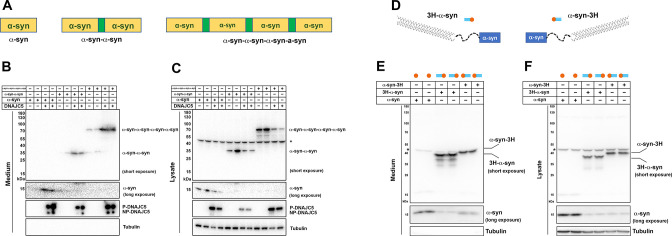
Figure 5—source data 1. Uncropped immunoblot corresponding to Figure 5.

Figure 5.

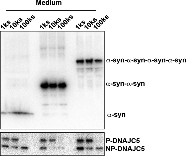
Figure 5—figure supplement 1. Medium fractionation of secreted tandem α-syn oligomers.

Figure 5—figure supplement 1.

Medium fractionation of secreted tandem α-syn oligomers. P-DNAJC5 was depleted in the supernatant after centrifugation at 100,000 (100k)×g. However, no significant decrease of tandem α-syn oligomers in the supernatant was observed.
Figure 5—figure supplement 1—source data 1. Uncropped immunoblot corresponding to Figure 5—figure supplement 1.
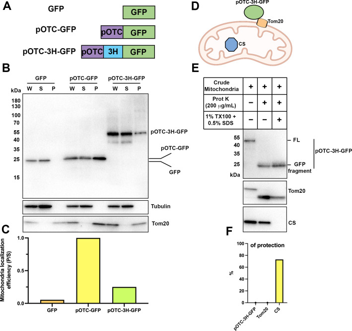
Figure 5—figure supplement 2. Thermostable three helix bundle blocked pOTC-mediated mitochondrial import of GFP.

Figure 5—figure supplement 2.

(A) Schematic diagrams of constructs used in mitochondrial import assay. GFP, GFP alone. pOTC-GFP, GFP with leader peptide from ornithine ranscarbamylase (OTC) fused at amino-terminus. pOTC-3H-GFP, the thermostable three helix bundle was inserted between the leader peptide pOTC and GFP. (B) Immunoblot analysis of proteins in crude mitochondria fraction. Whole-cell lysate (W) was prepared by mixing homogenized cells with equal volume of 2.3 M sucrose buffer and centrifugation at 1200×g to remove large debris. Soluble fraction (S) and particulate fraction (P) were separated by centrifuging W fraction at 7000×g for 10 min. Mitochondria were enriched in P fraction. Tom20, mitochondrial marker. Tubulin, soluble marker. (C) The mitochondrial localization efficiency was calculated by quantifying the protein amount in P fraction divided by the amount in S fraction. (D) Cartoon depicting relative localization of proteins inside or outside of mitochondria. Tom20, a mitochondrial outer membrane protein. Citrate synthase (CS), a mitochondrial matrix protein. The import of pOTC-3H-GFP is blocked by 3H. (E) Proteinase K protection assay of crude mitochondria. SDS (0.5%) was added in addition to 1% TX-100 to sensitize protease treatment of well-folded protein, for example, CS. The protease accessibility of pOTC-3H-GFP was revealed by the production of GFP fragment and disappearance of the full-length (FL) band. (F) Quantification of percentage of protection of proteins in (E).
Figure 5—figure supplement 2—source data 1. Uncropped immunoblot corresponding to Figure 5—figure supplement 2.