. 2023 Jan 10;12:e85837. doi: 10.7554/eLife.85837

Figure 6. XPACK (XP)-induced DNAJC5 L115R oligomerization rescued α-syn secretion.

(A) Ladder pattern of higher molecular weight (HMW) DNAJC5 oligomers in the medium. Medium from HEK293T cell culture transfected with DNAJC5 was centrifuged at 1000 (1k)×g, 10,000 (10k)×g, and 100,000 (100k)×g, followed by SDS-PAGE and immunoblot of supernatant (s) fractions at each centrifugation step. (B) Fractionation of HMW-DNAJC5 with gel filtration. HEK293T cells transfected with DNAJC5 were lysed, clarified, and subjected to gel filtration. HMW-DNAJC5 of different sizes were separated based on their corresponding molecular weight. (C) XP-DNAJC5 L115R mutant forms a membrane-bound oligomer. Cellular fractionation was performed with HEK293T cells transfected with indicated DNAJC5 variants. Note the substantial change of electrophoretic mobility of XP-DNAJC5 L115R on SDS-PAGE. (D) α-syn secretion induced by XP-DNAJC5 L115R. Secretion assay was performed with HEK293T cells transfected with indicated plasmids. About 10 μm 2-BA was used to block induced α-syn. (E) Expression of α-syn and DNAJC5 variants in HEK293T cells. Note the substantial change in electrophoretic mobility of 2-BA-treated XP-DNAJC5 L115R on SDS-PAGE.

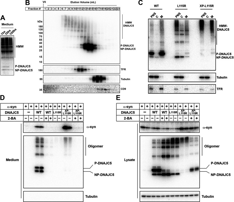
Figure 6—source data 1. Uncropped immunoblot corresponding to Figure 6.

Figure 6.

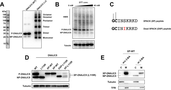
Figure 6—figure supplement 1. Characterization of HMW-DNAJC5 and XPACK fusion.

Figure 6—figure supplement 1.

(A) DNAJC5 (ΔJ) forms a series of SDS-resistant oligomers. HEK293T cells transfected with WT DNAJC5 or DNAJC5 (ΔJ) were lysed and evaluated by immunoblot using anti-DNAJC5 antibody. (B) HMW-DNAJC5 is not formed by non-specific disulfide bonds. HEK293T cells transfected with DNAJC5 were lysed for SDS-PAGE followed by immunoblot. The loading samples for SDS-PAGE were prepared with increasing amount of DTT up to 40 mM. (C) Schematic diagram of XPACK. XPACK is myristoylated on the first glycine (G) and palmitoylated on the second cystine (C). Replacement of Serine (S) at position 5 with a hydrophobic isoleucine (I) abolishes normal lipidation of XPACK. (D) Assessment of different XPACK fusion constructs. Cell lysate containing different DNAJC5 constructs were evaluated by anti-DNAJC5 immunoblot. (E) Membrane association of XP-DNAJC5 dependent on palmitoylation. HEK293T cells transfected with XP-DNAJC5 were treated with DMSO or 10 μM 2-BA. After cell culture for 24 hr, cellular fractionation was performed with homogenized cells. Distribution of XP-DNAJC5 in cytosol (C) and membrane (M) fractions were evaluated with immunoblot.
Figure 6—figure supplement 1—source data 1. Uncropped immunoblot corresponding to Figure 6—figure supplement 1.
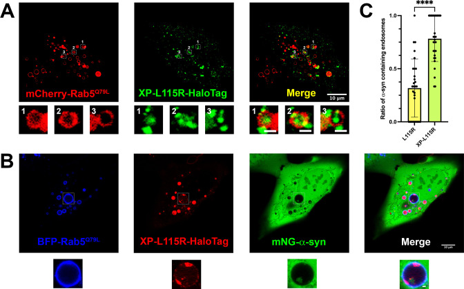
Figure 6—figure supplement 2. Live-cell images of U2OS cells expressing XP-DNAJC5 L115R mutant and α-syn.

Figure 6—figure supplement 2.

(A) Internalization of punctate XP-DNAJC5 (green) into enlarged endosomes labeled by mCherry-Rab5Q79L (red). U2OS cells were transfected with XP-DNAJC5 and mCherry-Rab5Q79L. Live-cell imaging was performed 24 hr after transfection. Representative enlarged endosomes with XP-DNAJC5 are shown in magnified inset. Scale bar: 10 μm in overviews and 1 μm in magnified insets. (B) α-syn enters into enlarged endosomes in the presence of XP-DNAJC5. XP-DNAJC5-HaloTag (red) and mNG-α-syn (green) were coexpressed in U2OS cells carrying BFP-Rab5Q79L (blue) mutant and imaged. Representative enlarged endosome with both XP-DNAJC5-HaloTag and mNG-α-syn inside is shown in magnified inset. Scale bar: 10 μm in overviews and 1 μm in magnified insets. (C) Quantification of the ratio of α-syn-containing endosomes in cells co-transfect with DNAJC5 L115R or cells co-transfected with DNAJC5 XP-L115R. More than 100 enlarged endosomes were counted in each group. Error bars represent standard deviations. P value<0.0001, two-tailed t test.