. 2023 Jan 10;12:e85837. doi: 10.7554/eLife.85837

Figure 7. Recapitulation of endogenous DNAJC5-mediated α-syn secretion in various neuronal cell cultures.

(A) Membrane and cytosol fractionation of differentiated SH-SY5Y neuroblastoma cells. The fractionation was performed as depicted in Figure 1B. C, cytosol; M, membrane. The distribution of endogenous DNAJC5 and α-syn was evaluated by immunoblot. Transferrin receptor (TFR) was used as a membrane marker. Tubulin was used as a cytosol marker. (B) Quantification of α-syn level in the supernatant of centrifuged media with ELISA. Conditioned media were collected and sequentially centrifuged at 1000 (1k)×g, 10,000 (10k)×g, and 100,000 (100k)×g. The supernatant from each centrifugation step (1ks, 10ks, and 100ks) was collected and measured by LEGEND MAX Human α-synuclein (Colorimetric) ELISA Kit. One-way ANOVA showed no significant (ns) difference of α-syn level between fractions. (C) Quercetin inhibited endogenous α-syn secretion in hiPSC-derived midbrain dopamine neurons. hiPSC-dopamine neurons carrying the GBA-N370S mutation were treated with quercetin (5 μM or 10 μM) at day 35. Culture media samples were harvested after 3 days treatment at day 38 and α-syn levels in the media were analyzed by electro-chemiluminescent immunoassay. Data points represent individual cell lines derived from different donors and are normalised to total protein in the corresponding cell lysates. One-way ANOVA followed by Tukey’s post hoc test shows a significant reduction in α-syn secretion with increasing quercetin concentration (*p<0.05, **p<0.01). (D) Depletion of endogenous DNAJC5 in SH-SY5Y cells decreased basal α-syn secretion. After 3 days of culture, the media from differentiated SH-SY5Y cells transduced with shRNA targeting GFP (shRNA-GFP) or shRNA targeting DNAJC5 (shRNA-DNAJC5) were collected and the extracellular α-syn was quantified with ELISA. P value<0.0002, two-tailed t test. (E) Expression of exogenous human DNAJC5 in mouse mDA stimulated basal α-syn secretion. WT mDA and mDA expressing hDNAJC5 were treated with DMSO or 100 nM BaFA1. Quantification of α-syn in conditioned media was performed with Mouse α-synuclein ELISA Kit (Abcam). α-syn secretion was normalized by dividing the α-syn in media (pg/ml) by the α-syn in cell lysates (ng/ml). P value<0.01, one-way ANOVA. (F) BaFA1 increased DNAJC5 oligomerization in mouse mDA neurons.

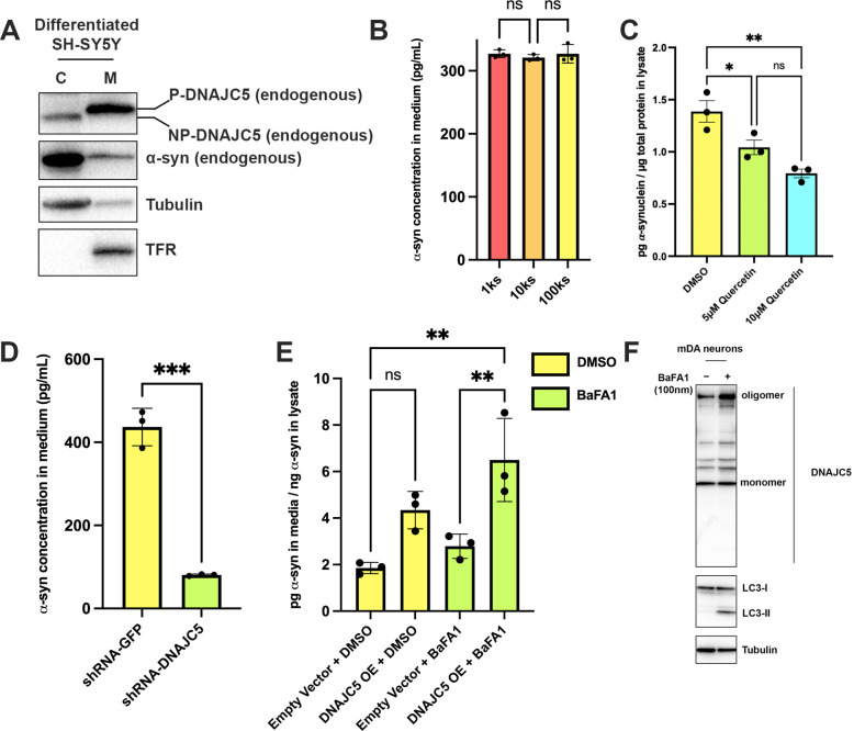
Figure 7—source data 1. Uncropped immunoblot corresponding to Figure 7.

Figure 7.

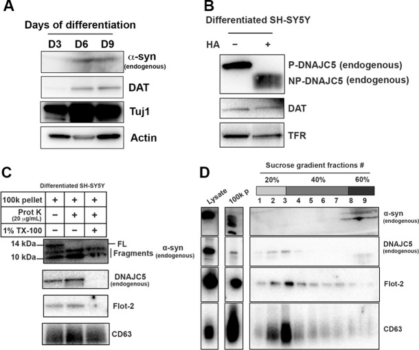
Figure 7—figure supplement 1. Basal α-syn secreted as a soluble form from differentiated SH-SY5Y.

Figure 7—figure supplement 1.

(A) Differentiation of SH-SY5Y was initiated by lowering the serum concentration to 1% FBS and addition of 10 µM retinoic acid (RA). Media were replaced every 3 days and the cells were harvested at indicated time to examine the expression of neuronal marker. DAT, dopamine transporter; Tuj1, neuron-specific class III β-tubulin. (B) In vitro depalmitoylation assay of endogenous DNAJC5 in differentiated SH-SY5Y cells. The depalmitoylation assay was performed as in Figure 1—figure supplement 1B using membrane (M) fraction from SH-SY5Y cells. HA, hydroxylamine. (C) Proteinase K protection assay of 100,000 (100k) pellet fraction from the centrifuged media of differentiated SH-SY5Y culture. Flot-2 and CD63 were used as exosome markers. (D) Sedimented α-syn was not buoyant. 100k pellet from (C) was mixed with 60% sucrose in PBS and layered with 40% and 20% sucrose in PBS sequentially. After centrifuged at 150,000×g for 16 hr, fractions were collected from top to bottom.
Figure 7—figure supplement 1—source data 1. Uncropped immunoblot corresponding to Figure 7—figure supplement 1.
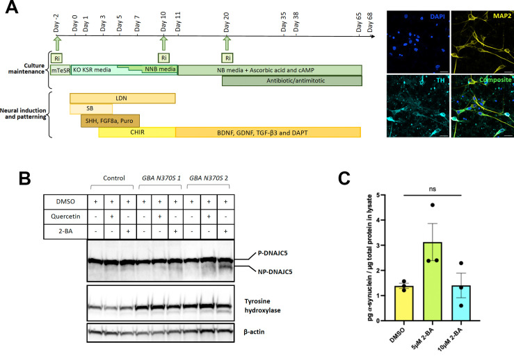
Figure 7—figure supplement 2. Differentiation of human induced pluripotent stem cells (hiPSCs) and palmitoylation of DNAJC5.

Figure 7—figure supplement 2.

(A) Schematic of the differentiation protocol used to generate hiPSC-derived dopamine neurons, including a patterning phase to generate neural progenitor cells followed by differentiation into mature neurons. Green arrows represent points of replating when cells are supplemented with ROCK inhibitor (Ri) to increase survival. BDNF, brain-derived neurotrophic factor; FGF8a, fibroblast growth factor 8a; GDNF, glial cell line-derived neurotrophic factor; Puro, puromorphamine; SHH, sonic hedgehog; TGF-β3, transforming growth factor beta 3 (see Kriks et al., 2011; Beevers et al., 2017; Lang et al., 2019 for full details). Neurons are mature and harvested between 35 and 68 days for analysis. Immunocytochemical fluorescent images of mature neurons at day 50 stained for neuronal marker microtubule-associated protein (MAP2), the dopaminergic marker tyrosine hydroxylase (TH), and DAPI. Scale bar: 20 µm. (B) DNAJC5 is palmitoylated in iPSC-derived dopamine neurons. Immunoblot analysis of DNAJC5 palmitoylation in hiPSC-derived dopamine neurons treated with DMSO, 7.5 μM Quercetin or 10 μM 2-BA on day 65 and harvested on day 68. TH was used as a marker of dopaminergic identity. (C) Partial depalmitoylation of DNAJC5 by 2-BA in iPSC-derived dopamine neurons does not reduce α-syn secretion. hiPSC-derived dopamine neurons carrying the GBA-N370S mutation were treated with palmitoylation inhibitor 2-BA (5 μM or 10 μM) at day 35. Culture media samples were harvested after 3 days treatment at day 38 and α-syn levels in the media were analyzed by electro-chemiluminescent immunoassay. Data points represent individual cell lines derived from different donors and are normalized to total protein in the corresponding cell lysates. One-way ANOVA shows no effect of 2-BA on α-syn secretion.
Figure 7—figure supplement 2—source data 1. Uncropped immunoblot corresponding to Figure 7—figure supplement 2.
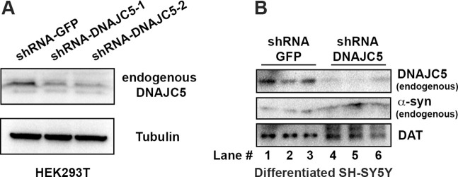
Figure 7—figure supplement 3. shRNA-mediated DNAJC5 knockdown in differentiated SH-SY5Y cells.

Figure 7—figure supplement 3.

(A) Examination of knockdown efficiency by shRNA targeting DNAJC5 in HEK293T cells. (B) Endogenous DNAJC5 expression decreased in SH-SY5Y cells transduced with shRNA targeting DNAJC5.
Figure 7—figure supplement 3—source data 1. Uncropped immunoblot corresponding to Figure 7—figure supplement 3.
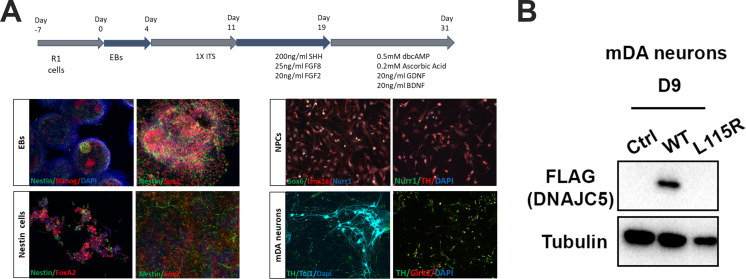
Figure 7—figure supplement 4. Differentiation of mouse embryonic stem cells (mESCs) and expression of human DNAJC5.

Figure 7—figure supplement 4.

(A) Schematic overview of protocol used for differentiation of mESCs into middle-brain dopaminergic (mDA) neuronal cultures. Immunocytochemical staining using stem cell markers (Nanog, Sox2), neuronal precursor marker (Nestin), mDA markers [FoxA2, Nurr1, Lmx1a, Sox6 (selective marker of substantia nigra pars compacta lineage), TH (tyrosine hydroxylase), Girk2 (G-protein-regulated inward-rectifier potassium channel 2, expressed in DA neurons)], neuronal marker (Tuj1) and nuclear marker (dapi). (B) Expression of human DNAJC5 with C-terminal FLAG tag in mDA neurons.
Figure 7—figure supplement 4—source data 1. Uncropped immunoblot corresponding to Figure 7—figure supplement 4.