Figure 6.
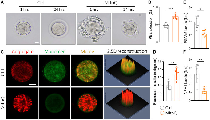
Effect of MitoQ on in vitro maturation of aging mouse oocytes and mitochondrial function. All oocytes were matured in vitro with 10 nM MitoQ for 24 h from 51-week-old B6 mice. (A) Representative images of in vitro maturation of oocytes from aged mice. (B) The quantification of first polar body extrusion from mice oocytes. (C) Mitochondrial membrane potential (Δψm) assessed by JC-1 staining in control and supplemented MitoQ oocytes (red, Δψm high; green, Δψm low). (D) Quantification of the ratio of red to green fluorescence intensity in control and MitoQ-supplemented oocytes. (E, F) The levels of oxeiptotic core genes were determined by qPCR. Scale bar, 25 μm. *p < 0.05, **p < 0.01, ***p < 0.001.

