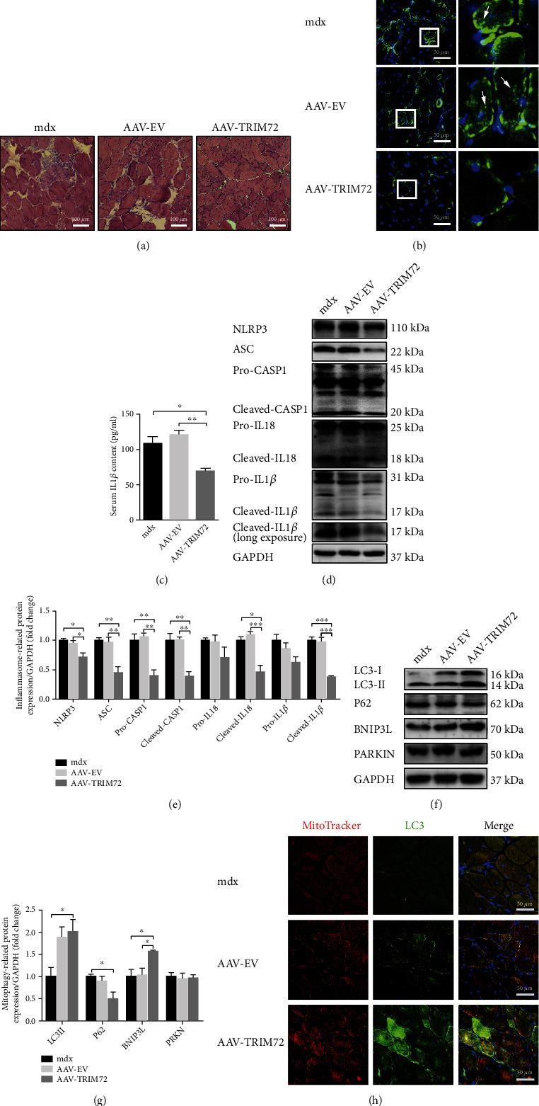
Figure 2.

Decreased NLRP3 inflammasomes and increased mitophagy in AAV-TRIM72 mdx mice. (a) Representative HE staining images of tibial anterior muscles in mdx mice with or without AAV transfection. Scale bar, 100 μm. (b) Immunofluorescence was performed with specific antibody targeting NLRP3. As shown by arrows in the magnification inset, NLRP3 dots were reduced in AAV-TRIM72 mdx mice. Scale bar, 50 μm. (c) Serum IL-1β level detected by ELISA assay, n = 4. (d, e) Western blot analysis and quantification of NLRP3, ASC, IL18, caspase-1, and IL-1β in three mdx groups, n = 4. (f, g) Immunoblot analysis and quantification of P62, LC3, BNIP3, and PARKIN in lysates of three mdx groups, n = 4. (h) Representative immunofluorescence images of LC3, MitoTracker, and the merged images in three mdx groups. Scale bar, 50 μm. Data were expressed as mean ± SEM. ∗p < 0.05, ∗∗p < 0.01.
