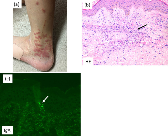
Figure 1.
Skin lesion. a: Purpura on the lower leg. b: Light microscopy: Neutrophils with nuclear debris and eosinophils (arrow) are seen surrounding blood vessels in the subcutaneous tissue (Hematoxylin and Eosin staining). c: Immunofluorescence microscopy: Blood vessels showed positivity for IgA (arrow).

