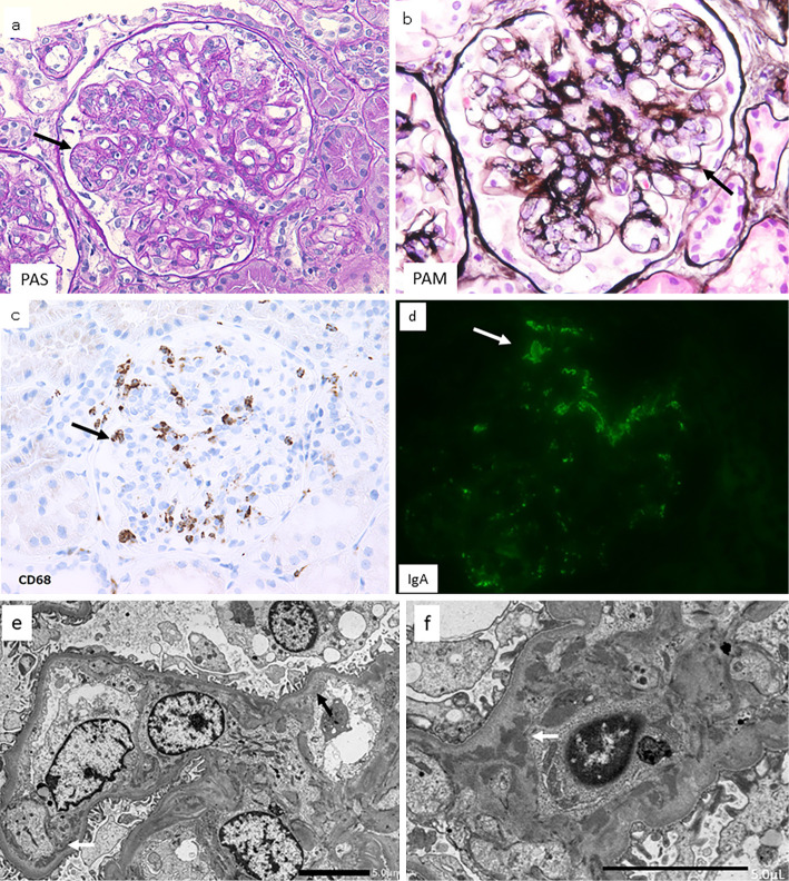
Figure 2.
Kidney lesion. Microscopy findings of the kidney biopsy specimen. a: Light microscopy: Almost all glomeruli displayed endocapillary glomerulonephritis that was characterized by endothelial cell swelling (arrow), resulting in the obstruction and/or narrowing of the glomerular cavity; periodic acid-Schiff staining. b: Light microscopy: Double-contour appearance and thickening of the glomerular basement membrane (arrow) (periodic acid-methenamine silver staining). c: Light microscopy: CD68-positive cells (arrow) were seen in the glomeruli (CD68 staining). d: Immunofluorescence microscopy: Glomeruli showed mild positivity for IgA (green area, arrow). e: Electron microscopy: Endothelial cells were swollen, and subendothelial edema was seen (black arrow); electron-dense deposits (white arrow) were found in the subendothelial space. f: Electron microscopy: electron-dense deposits (white arrow) were found in the paramesangial area.

