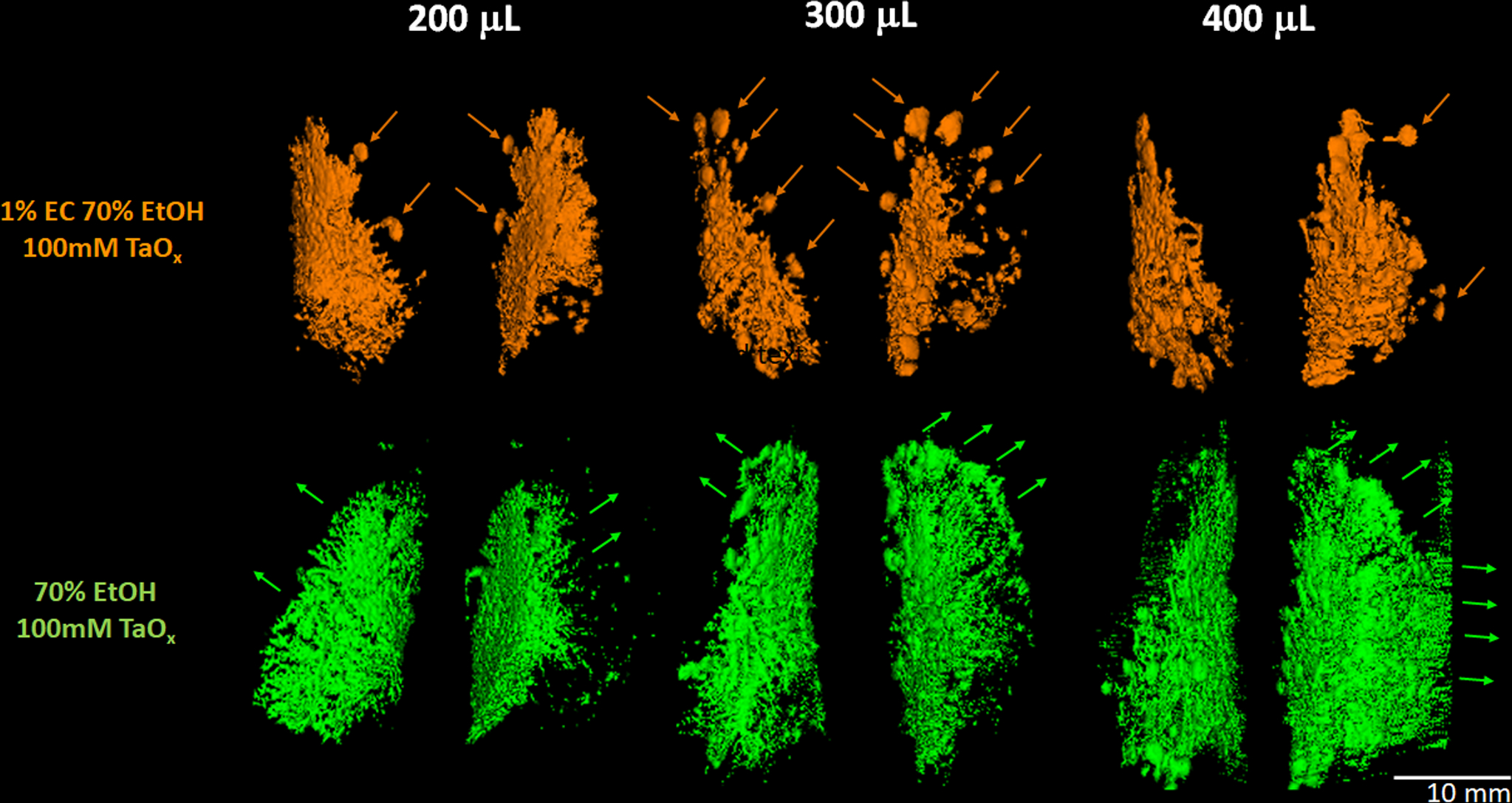
Figure 3. 3D reconstruction and assessment of ablative solution filling and diffusion.

70% EtOH/100 mM TaOx nanoparticles with 1% EC (top) or without EC (bottom) were intraductally injected into the second abdominal mammary gland pair (#4 and #10) and immediately imaged by micro-CT. Each Sprague-Dawley rat received an increasing volume of either solution. Individual ductal trees were reconstructed using an image analysis software package (spline trace + propagate object + threshold rendition). With 1% EC, the solution can be seen reaching the terminal ends. As delivered volume is increased the number of TEBs filled is more apparent. Scale bar corresponds to 10 mm in all renditions.
