
Figure 12. Aortic Imaging Techniques to Determine the Presence and Progression of Aortic Disease.
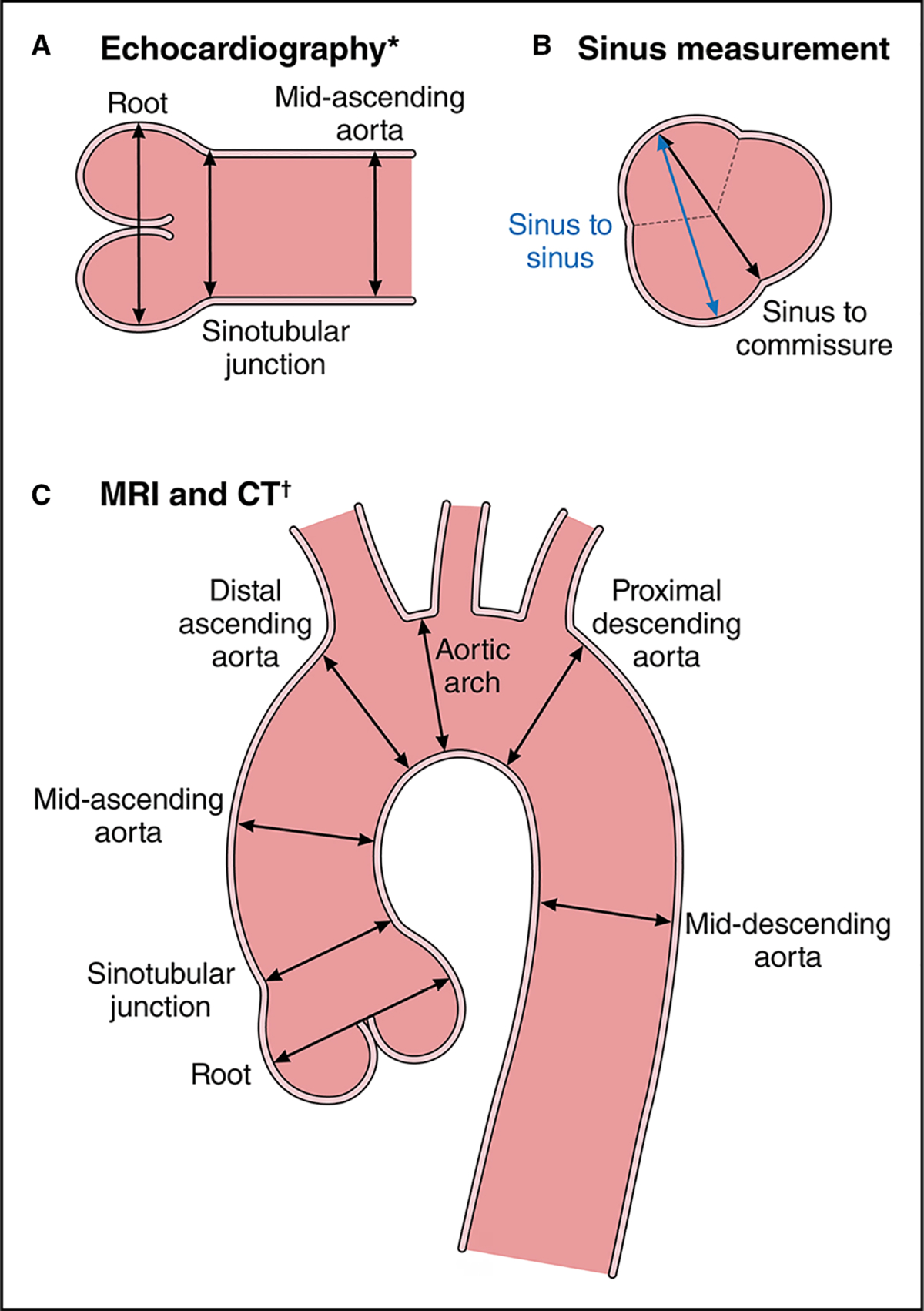
(A) Schematic shows the leading-edge to leading-edge measurement technique used in echocardiography, from left to right: measurement of the aortic root (sinuses of Valsalva), sinotubular junction, and proximal tubular ascending aorta. (B) Inner-wall to inner-wall measurements of the aortic root used in MRI and CT. In addition, a consistent approach to measuring all 3 sinuses with MRI and CT is necessary. The sinus-to-commissure and sinus-to-sinus measurements can both be used, but consistency is necessary for interval surveillance. (C) Standard measurement locations for MRI and CT with the inner-wall to inner-wall technique. Adapted from Borger et al.21 Copyright 2018, with permission from Elsevier, Inc. CT indicates computed tomography; and MRI, magnetic resonance imaging. *Leading-edge to leading-edge. †Inner-wall to inner-wall.