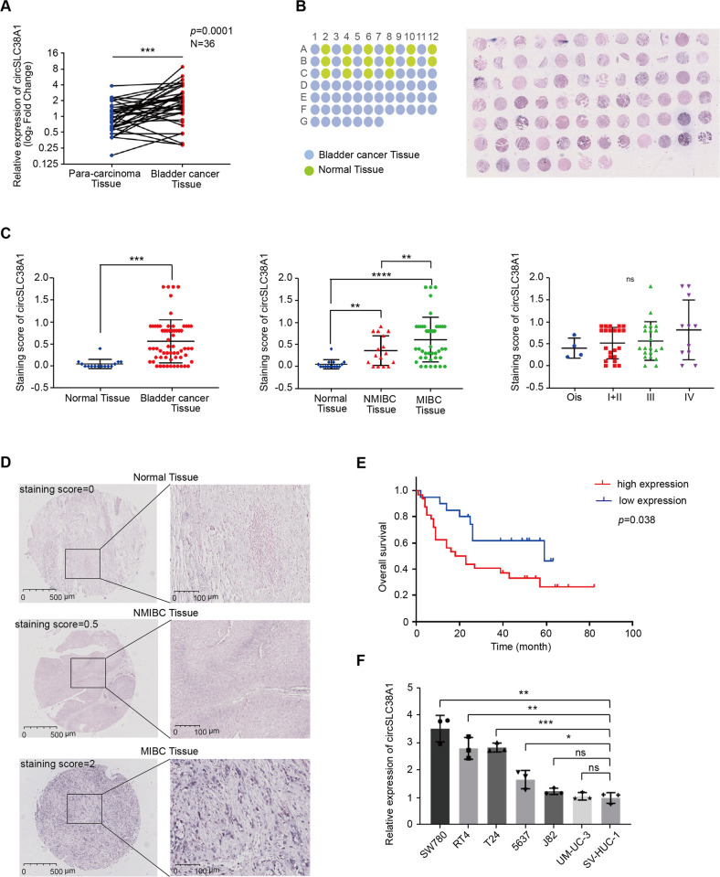
Fig. 2. circSLC38A1 is upregulated in BC.
A Expression levels of circSLC38A1 in 36 pairs of BC and adjacent normal tissues were detected by the divergent primers (P = 0.0001). B The expression level of circSLC38A1 was measured by RNA ISH staining tissue microarrays (n = 79). C The difference in staining score between normal tissues and BC tissues (left), the difference in staining score between NMIBC tissues and MIBC tissues (middle), and the correlation of circSCL38A1 expression with tumor node metastasis classification (TNM) of bladder cancer (left). The staining score = staining intensity score * staining positive rate. (staining intensity score 0 is defined as negative, staining intensity score 1+ is defined as weak expression, staining intensity score 2+ is defined as moderate expression, and staining intensity score 3+ is defined as a strong expression). D The representative staining images were shown. E Kaplan–Meier analysis of the correlation between circSLC38A1 expression and overall survival. Patients with high levels of circSLC38A1 had significantly shorter overall survival (P = 0.038). The P value was determined by a Log-rank test. F Relative expression of circSLC38A1 in BC cell lines and human normal urothelial cell line measured by qRT-PCR. Data are presented as means ± standard deviation from three independent experiments. *p < 0.05, **p < 0.01, ***p < 0.001, ****p < 0.0001.

