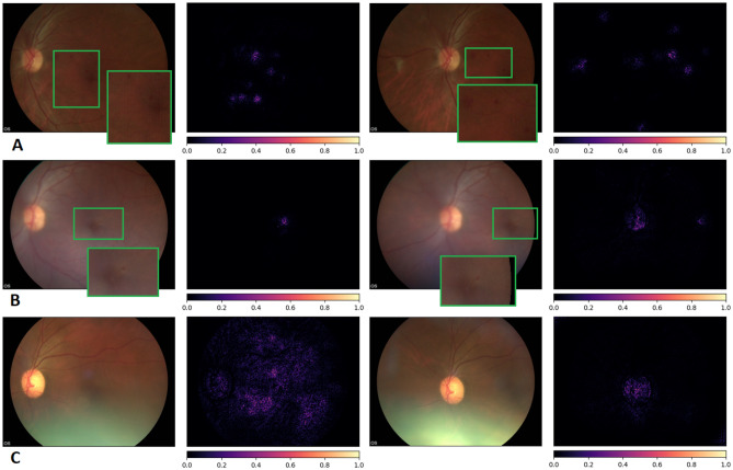
Figure 4.
Integrated gradients pixel attributions. Pairs of macula and optic disc centred images of three different patient eyes and the obtained saliency maps (A) Severe NPDR, referable DME, DLS score 0.98. (B) Moderate NPDR, No DME, DLS score 0.58, (C) No DR, No DME, 0.04. The saliency maps highlight relevant lesions for the diagnosis (microaneurysms). In the absence of referable DR/DME, only the optic disc or general regions of the retina are highlighted.

