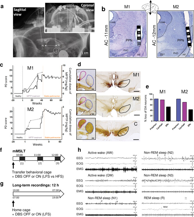
Fig. 1. Surgery report, PD scores and experimental design.
a X-ray of the final implantation of the electrode in the PPN area for M1, in both coronal and sagittal view with the internal landmarks AC-PC line determined by ventriculography (LV: lateral ventricle). b Micrographs of Cresyl-violet stained sections through the PPN of M1 and M2 showing postmortem reconstruction of the electrode track and position of the contacts, (-) is the active contact. CGMB = central gray substance of midbrain; PnO= oral pontine reticular nucleus; ml= medial lemniscus. c Graphs illustrating the longitudinal progression of parkinsonian syndrome for M1 and M2, induced by injection of chronic low doses of MPTP, based on weekly observations. The solid black line shows the PD score and the gray dotted line represents the cumulative dose of MPTP with each dot corresponding to an injection of 0.2–0.5 mg/kg. Note 3 key periods: the healthy period (black), the MPTP treatment period (gray) and the stable parkinsonian period (orange). During the healthy period the PD scores were 0/25. During the stable parkinsonian period the score was 13.8 ± 0.2 / 25 for M1 and 18.9 ± 0.1 / 25 for M2. d Micrographs showing tyrosine hydroxylase (TH) immunostaining, at the level of the striatum (framed image, scale bar= 2000µm) and the substantia nigra (scale bar= 2000µm) for M1, M2 and the control animal (C). The circled areas correspond to the striatal structures (putamen in blue and caudate nucleus in purple) and the substantia nigra in black. e Graphs showing the percent loss of TH expression in the putamen, the caudate nucleus and the substantia nigra of M1 and M2 compared to the control animal. Percent loss are calculated based on optical density method using the region of interest outlined in d. f Design of the modified multiple sleep latency test (mMSLT) with 20 min light-OFF at 10:00 h (1), 11:00 h (2) and 12:00 h (3), performed in behavioral cage and under different conditions: healthy or stable parkinsonian states with PPN-DBS OFF or ON (LFS vs. HFS). g Design of long-term recordings of 12 h nighttime (from 19:00 h to 7:00 h) and daytime (from 7:00 h to 19:00 h), performed in home cage and under different conditions: healthy or stable parkinsonian states with PPN-DBS OFF or ON (LFS only). h Polysomnographic recordings for wake/sleep stages analysis. Thirty seconds epochs showing active wake (AW), quiet wake (QW), non-REM sleep stage 1 (N1), stage 2 (N2), stage 3 (N3) and REM sleep (R).

