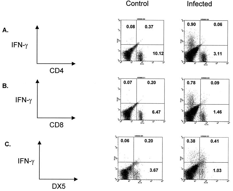
FIG. 2.
Intracellular cytokine staining of IFN-γ in lung digest cells. On day 2 after i.t. challenge with L. pneumophila (i.e., when IFN-γ levels were at their peak), mice were sacrificed to obtain cells for lung digest. The cells were stained for surface expression of CD4 (A), CD8 (B), or DX5 (C) and then costained for intracytoplasmic IFN-γ. After staining, cells underwent analysis by flow cytometry with gating for lymphocytes by size and complexity characteristics (41). (C) The majority of cells staining positive for intracellular IFN-γ were DX5+, although a number of IFN-γ-positive cells were DX5−, CD4−, and CD8−. The experiment was performed twice (n = 2 animals per group).

