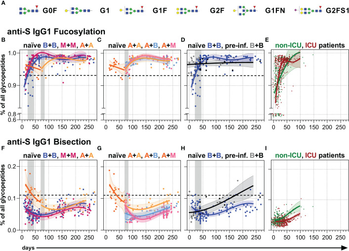
Figure 4.
Anti-S serum IgG1 fucosylation and bisection. (A) The six major IgG Fc N-glycans attached to Asn 297 of IgG1 with an average relative abundance of more than 3% ( Table S2 ): Galactose: G, yellow circle; sialic acid: S, purple diamond; fucose: F, red triangle; mannose: green circle; N-acetylglucosamine: GlcNAc and bisecting GlcNAc, N, blue square. (B–D) Anti-S serum IgG1 Fc N-fucosylation and (F–H) anti-S serum IgG1 Fc N-bisection of the indicated six vaccination groups. The used color codes are identical to Figure 1D . Gray bars: time windows of the second shot after the first shot with an mRNA (between day 21 and 45) or adenovirus-based (between day 70 and 84) vaccine. Dashed lines indicate the average level of total IgG1 Fc fucosylation or bisection, respectively ( Figure S6 ). (E) Anti-S serum IgG1 Fc N-fucosylation and (I) anti-S serum IgG1 Fc N-bisection of unvaccinated, hospitalized non-ICU and ICU SARS-CoV-2 patients from our previous study (60) for comparison.

