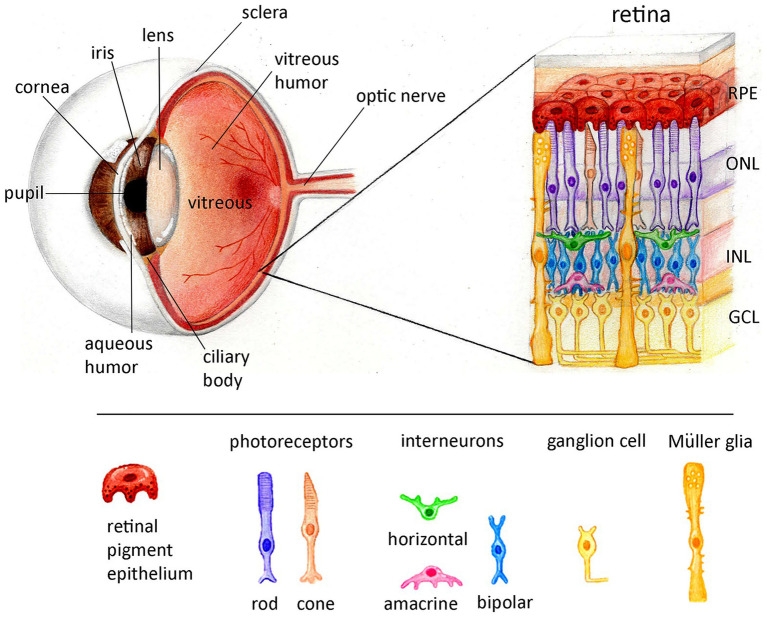
Figure 1.
Schematic representation of the anatomical features of the eye and the lamination of the retina. The eye is anatomically divided in three layers. The external layer of the eye consists of the cornea, and sclera. The intermediate layer includes-at the anterior-the iris and ciliary body and in the posterior the choroid (not shown here). The internal layer is comprised by the retina. The space is filled with fluid called the aqueous humor, at the anterior chamber, or vitreous humor between the lens and the retina. The retina is divided in two compartments: one non-neuronal, the retina pigment epithelium (RPE) and one neuronal, the neuroretina. The vertebrate neuroretina is laminated in three layers of nerve cell bodies and two layers of synapses. The outer nuclear layer (ONL) consists of the somata of the rods and cones, the inner nuclear layer (INL) consists of the cell bodies of the bipolar, horizontal and amacrine interneurons and the ganglion cell layer (GCL) contains cell bodies of ganglion cells and some displaced amacrine cells. The axons of the ganglion cells are forming the optic nerve interconnecting the retina with the brain.

