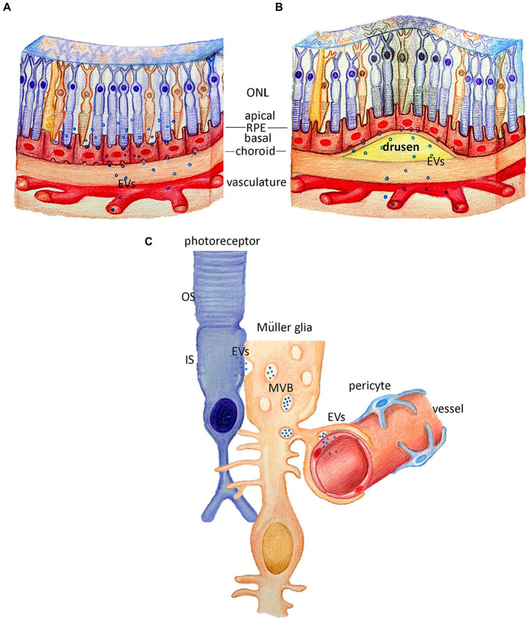
Figure 3.
Potential roles of extracellular vesicles in retina pigment epithelium and Müller Glia cells. (A) Schematic representation of the retina, where retina pigment epithelium (RPE) is potentially releasing extracellular vesicles (EVs) from the apical and basal side under healthy conditions. (B) Schematic representation of the retina with drusen formation, where the RPE EV release from the basal side, may potentially contribute to the drusen pathology. (C) Schematic representation of the suggested roles of the EVs release by the Müller glia cells, either via MVB release of direct budding, under healthy conditions where the EVs can be released towards photoreceptors or towards the vascular endothelium.

