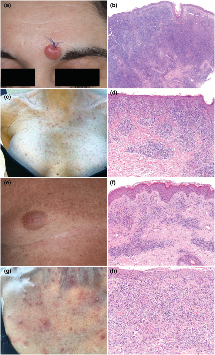
FIGURE 1.

(a) A single erythematous nodule on the forehead. (b) A diffuse dense infiltrate in the dermis, sparing the epidermis and composed of small‐ to medium‐sized hyperchromatic lymphocytes. Atypical lymphocytes, arranged in clusters, are CD4+, PD1+ and exceptionally GATA3+ (data not shown) consistent with primary cutaneous CD4+ small/medium T‐cell lymphoproliferative disorder [haematoxylin and eosin (H&E), 10×]. (c) an erythematous maculopapular eruption on the chest. (d) Histopathological findings diagnostic for atypic pytiriasis lichenoides and varioliformis acuta: A dense lichenoid and perivascular infiltrate in the upper dermis composed of small‐medium sized, hyperchromatic lymphocytes mixed with some blasts, few plasma cells, mast cells and red blood cells. Lymphocytes are positive for CD3, CD4, CD7 (H&E 20×), Ki‐67 20% (data not shown). (e) A diffuse and confluent erythematous papular eruption on the trunk. (f) Histology revealing lymphomatoid papulosis type A: Perivascular infiltrate in the superficial dermis of medium/large‐sized pleomorphic lymphocytes mixed with eosinophils and blast cells (around 30% of the infiltrate). Atypical lymphocytes were CD3+, CD4+, CD7+, CD30+ (blasts included) and GATA3+ (data not shown) (H&E 20×). (g) A diffuse vesicular and papulo‐pustular eruption on the chest. (h) A dense perivascular and interstitial infiltrate in the superficial dermis of small‐sized and large, blast‐like, pleomorphic lymphocytes mixed with numerous neutrophils, eosinophils and histiocytes. Large cells were CD3+, CD4+, CD7+, CD30+, GATA3+ (data not shown) consistent with lymphomatoid papulosis type A (H&E 20×).
