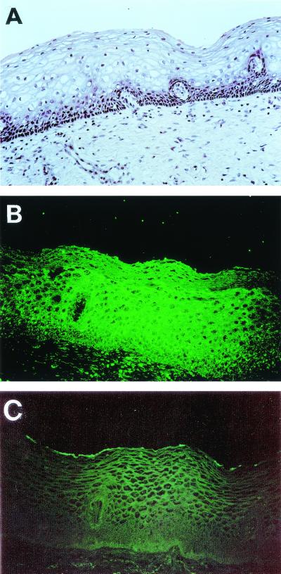
FIG. 3.
In situ assay of binding of S-pilin to fixed sections of human cervical tissue. (A) Tissue section stained with hematoxylin-eosin. (B) Tissue section overlaid with purified S-pilin preparation of MS11-8 (1.5 μg/ml). (C) Tissue section overlaid with concentrated supernatant of the P−n variant (20 μg/ml). Bound S-pilin was detected with PilE antiserum and FITC-conjugated anti-rabbit IgG. The low fluorescence level still detected represents the background of the FITC-labeled secondary antibody.

