Figure 1.

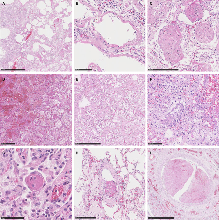
Histology of lung tissue of severe COVID‐19 and influenza. An overview of COVID‐19 lung tissue (A) displays features of acute DAD consisting of intra‐alveolar dispositions of hyaline membranes (B) and scattered ‘balls’ of fibrin indicative for AFOP (C). Lung tissue of influenza samples show features of acute and organising DAD. Extensive intra‐alveolar haemorrhage and macrophages (D) are indicative for acute DAD, while patchy distribution of fibroblastic proliferations (E) and squamous metaplasia (F) are features fitting organising DAD. Displayed features indicative for acute DAD, organising DAD and AFOP were interchangeable between COVID‐19 and influenza. G–I, Thrombi of various sizes from COVID‐19 samples. Microthrombi (G) as well as thrombi in small or large arteries (H, I) were more often seen in COVID‐19 than influenza. AFOP, acute fibrinous and organising pneumonia; DAD, diffuse alveolar damage.
