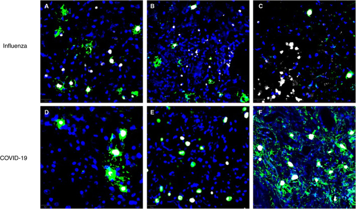
Figure 6.

Examples of multiplex immunofluorescence from different influenza (A–C) and COVID‐19 (D–F) tissue samples. Tryptase and chymase are stained green and white, respectively; nuclei are stained blue by 4′,6‐diamidino‐2‐phenylindole (DAPI). Note that the vast majority of tryptase‐positive cells in COVID‐19 samples are also positive for chymase, while a substantial amount of tryptase‐positive cells are negative for tryptase in influenza samples. [Color figure can be viewed at wileyonlinelibrary.com]
