
Figure EV5. Structural comparison of antibody‐binding epitope and amino acid conservation in the RBD.
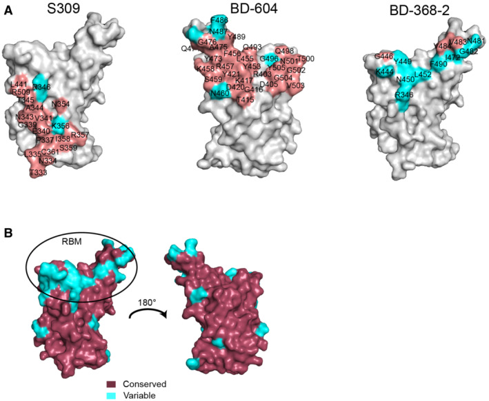
- Comparison of the binding interface between the SARS‐CoV‐2 antibody and the RshSTT182/200 RBD. In S309 (PDB: 7R6X), BD‐604, and BD‐368‐2 (PDB: 7CHF) binding to the SARS‐CoV‐2 RBD site, the conserved residues between the SARS‐CoV‐2 and RshSTT182/200 RBDs are shown in salmon, while variable residues are shown in cyan.
- Conserved residues and variable residues between the SARS‐CoV‐2 and RshSTT182/200 RBDs are shown in raspberry and cyan, respectively.