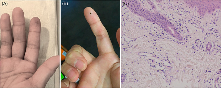
FIGURE 1.

The pathology and therapeutic outcome of fingertip lesion before and after treatment. The right index fingertip showed cyanosis before treatment (A). (B) denotes a fully recovery of cyanosis with a post‐biopsy wound after 25 days follow‐up with treatment. Pathology of cyanotic fingertip showed dilated vessels with scanty inflammatory infiltrates (C).
