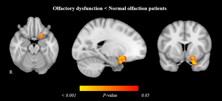
Figure 1.

Gray matter volume reduction in olfactory dysfunction patients compared with normal olfaction COVID‐19 patients. Significant voxel‐wise difference is marked in warm colors. Results are displayed over the sagittal, coronal, and axial sections of the MNI 152 standard brain at p‐value ≤0.05 FWE‐corrected. MNI, Montreal Neurosciences Institute; R, right.
