FIG. 1.
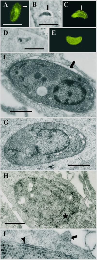
Early intracellular development (1 h to 2 days postinfection) of sporozoites of S. singaporensis in L2 rat pneumonocytes in vitro. Immunofluorescence images, corresponding phase-contrast images, and ultrastructural morphology are shown. Bars, 1 μm (F to I) or 10 μm (A to E). (A) Immunofluorescent staining of a sporozoite 2 h postinfection using rabbit anti-sporozoite antibodies (K3). The banana-shaped sporozoite is located inside a PV, and the PVM (arrow) is labeled due to incorporated parasite molecules. The label of the PVM persisted up to 18 h postinfection but was reduced or absent at later intervals. (B) Phase-contrast image of a sporozoite which appears to leave the PV and enter the cytoplasm of the host cell (2 h postinfection). The arrow indicates the PV, which is clearly visible as a halo surrounding the parasite. Note the slender appearance and dark cytoplasm of the zoite. (C) Corresponding immunofluorescent staining with K3 antibodies. The arrow points at the PVM, which seems to be absent at the left (apical) end of the sporozoite (the nucleus is located closer to the posterior end; see panels F and G), possibly a sign of a disrupted PVM. Note that the K3 antibodies preferentially label the apical portion of this stage. (D) Phase-contrast image of a sporozoite located inside the cytosol; these stages assumed a stumpy appearance and the parasite's cytoplasm became lucent. (E) Corresponding immunofluorescence showing that the K3 label is evenly distributed throughout the cytoplasm. (F) Ultrastructure of a sporozoite 1 h postinfection. The zoite resides in a PV (arrow). Note the parasite's electron-dense cytoplasm and remnants of membranes adhering to the pellicle, indicating recent invasion of the cell. (G) Ultrastructure of a sporozoite residing free in the cytoplasm 2 h postinfection. (H) Late sporozoite/young schizont 2 days postinfection. Note the absence of rhoptries and micronemes, the enlarged nucleus, and formation of islets of granules of the crystalloid body (asterisk). (I) Enlarged view of the host-parasite interface of the previous specimen. The schizont resides inside the cytosol; the arrowhead indicates the pellicle, and the arrow indicates the plasmalemma of the host cell.

