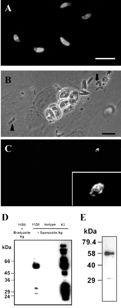
FIG. 6.
Characterization of MAb 11D5/H3 by indirect immunofluorescence and immunoprecipitation of the target antigen. Bars, 10 μm. (A) Staining pattern of air-dried sporozoites when the antibody was applied after the permeabilization of parasites with acetone. (B and C) Phase-contrast image and corresponding immunofluorescence of L2 pneumonocytes ethanol-acetone-fixed 1 h postinfection with sporozoites. Sporozoites were preincubated with mouse MAb 11D5/H3 before inoculation onto cells. After fixation and permeabilization of cells, 11D5/H3 was visualized with FITC-conjugated anti-mouse IgG. An extracellular sporozoite (arrow; above the focus level of host cell cy- toplasm) is labeled on the surface (inset shows a magnification) as indicated by the patchiness of membrane staining, whereas the label is absent on an intracellular sporozoite (arrowhead). The latter resides inside a PV which is visible by the surrounding halo. (D) Electrophoretic separation of the antigen precipitated by MAb 11D5/H3 from sporocyst-sporozoite extracts in gel-loading buffer containing 4% 2-mercaptoethanol (lane 2). A major band at 58 kDa was detected, whereby two minor bands appeared at 27 and 31 kDa. MAb 11D5/H3 did not react with bradyzoite extracts (lane 1), indicating that the antigen was not expressed in bradyzoites. An irrelevant isotype-matched MAb (IgG2a) and a sporozoite-specific rabbit serum (K3) served as negative and positive controls, respectively. (E) Electrophoretic separation of the antigen precipitated by 11D5/H3 from sporocyst-sporozoite extracts as performed before, except that reducing conditions were lowered to 2% 2-mercaptoethanol.

