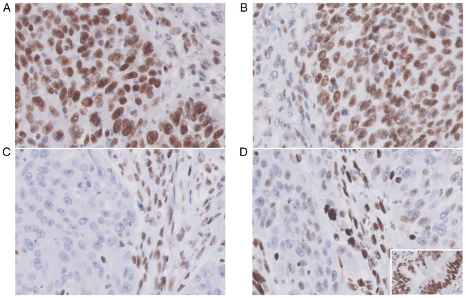
Figure 4.
MMR protein expression patterns showed positive nuclear staining (MMR-proficient) of (A) MLH1 protein (anti-MLH1 ES05, 400×) and (B) PSM2 protein (anti-PSM2 EP51); (C) MSH2 nuclear staining was totally negative (MMR-deficient) (anti-MSH2 FE11, 400×); (D) The staining pattern for MSH6 immunoexpression was heterogeneous (anti-MSH6 EP49, 400×) with only a partial loss of nuclear staining; (D, inset) The external control was positive in the benign glandular epithelium; (A-D) The internal control was positive in lymphocytes, stroma and endothelial cells for each MMR protein staining.

