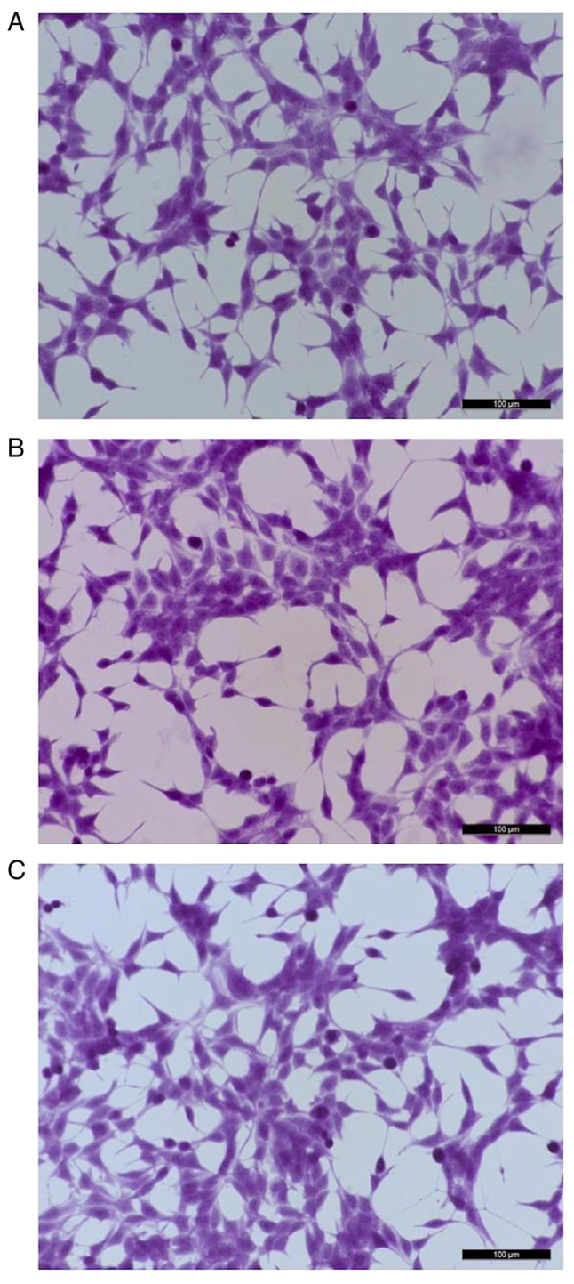
Figure 1.

Representative images of LNCaP cells stained with hematoxylin-eosin. (A) Control (untreated LNCaP cells). (B) LNCaP cells after exposure to 1×10⁷ pfu/ml of bacteriophage MS2 for 24 h. (C) LNCaP cells after exposure to 1×10⁷ pfu/ml bacteriophage MS2 for 48 h. No marked morphological differences were observed between the treated and untreated cells even after 48 h of exposure to bacteriophage MS2. Scale bars, 100 µm.
