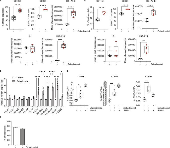
Fig. 3. Zabadinostat augments MHC expression in dendritic cells, and activates T and B lymphocytes.
a Flow cytometry analysis of the extracellular MHC class II protein level in bone marrow (collected from C57BL/6)-derived dendritic cells (CD11c+/CD86+) treated with 1 µM zabadinostat or DMSO control for 48 h; n = 5, Student’s t test; the acetylation mark (H3AcK14) and total histone 3 level were detected by flow cytometry; n = 5, Student’s t test. b Quantitative reverse transcription PCR (qRT-PCR) of MHC class I and II genes was also performed; n = 5; results presented as mean values +/−SD; one-way ANOVA. c Flow cytometry analysis of the extracellular MHC class II protein level in bone marrow (collected from Balb/c)-derived dendritic cells (CD11c+/CD86+) treated with 1 µM zabadinostat or DMSO control for 48 h, n = 5; the acetylation mark (H3AcK14) and total histone 3 level were detected by flow cytometry; n = 5; Student’s t test. d Activation of CD4, CD8 T cells and B cells upon 1 µM zabadinostat treatment or DMSO control was evaluated in splenocytes collected from Balb/c mice; n = 5; one-way ANOVA. e Viability of splenocytes measured by flow cytometry with L/D staining; graph represents pooled results from two animal experiments (see Fig. 5 (n = 4) and Supplementary Fig. 15 (n = 4)); results presented as mean values +/−SD.

