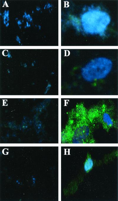
FIG. 3.
Fluorescence micrographs of B. henselae GFP reporter constructs. Panels A, C, E, and G show B. henselae GFP reporter constructs cultured in media alone. Panels B, D, F, and H show B. henselae GFP reporter constructs cocultured with HMEC-1. Reporter constructs shown are B. henselae pANT3/882Str (A and B), B. henselae pVBGFPR/882Str (C and D), B. henselae pVBGFPF/882Str (E and F), and B. henselae pVBDEL/882Str (G and H). Host cell and bacterial DNA is stained with DAPI. Overall magnification in each panel is ×400.

