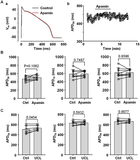
Fig. 2. Effect of SK channel blockers on APD.
A: (a) Overlay of APs recorded in control and after application of SK channel blocker apamin (100 nM). (b) Time course of changes of APD50 during application of apamin recorded from the same myocyte as in panel Aa.
B: APD50, APD70 and APD90 in control and in the presence of apamin (n/N=9/3).
C: APD50, APD70 and APD90 in control and in the presence of 1 μM UCL1684 (n/N=6/3).
Statistical analysis was performed using paired t test.

