Figure 6.
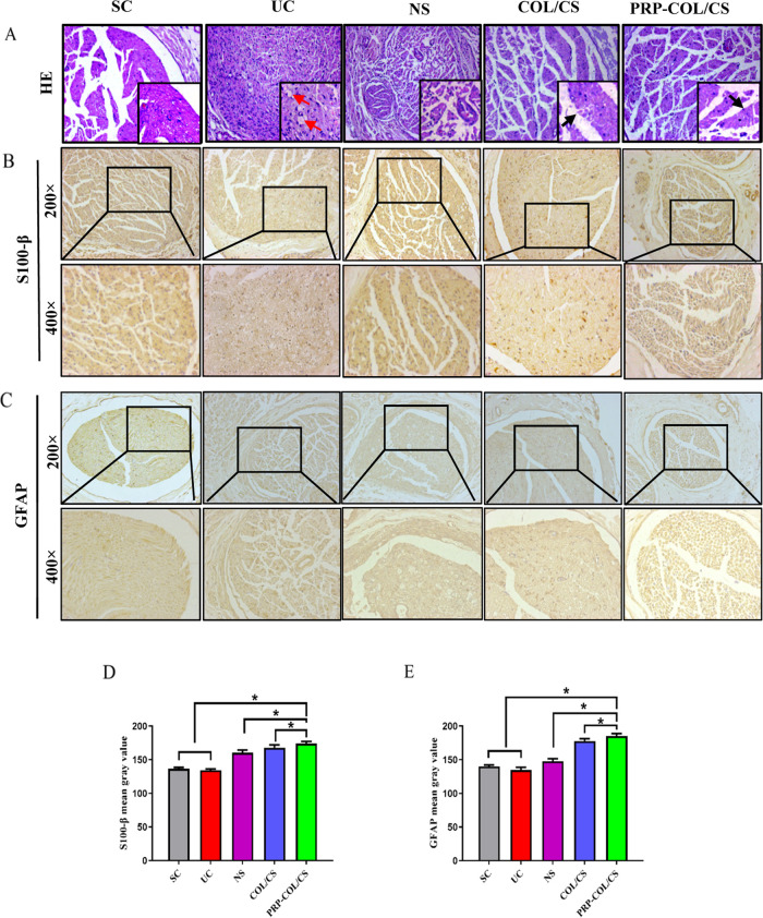
COL/CS composite film improved nerve morphology following SNI in rats. (A) Micrographs of the sciatic nerve stained by hematoxylin and eosin (H&E) at 8 weeks after nerve injury (×200 magnification and bottom right is ×400 magnification). The red arrow indicates nerve fiber vacuolation; the black arrow indicates myelin sheath. Immunohistochemical analysis of (B) S-100β and (C) GFAP. (D) Quantitative analysis of the S-100β mean gray value. (E) Quantitative analysis of the GFAP mean gray value. Values represent the mean ± SD, n = 5, *p < 0.05.

