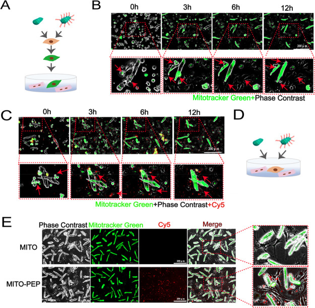
Figure 7.
Translocation mechanism of mitochondria into cardiomyocytes. (A) Schematic representation of the coculture strategy of murine endothelial cell line (bEnd.3) with cardiomyocytes. After coincubated with mitochondria, the bEnd.3 were cocultured with cardiomyocytes. (B,C) Living cell imaging detection of dynamic changes of transplanted mitochondria or PEP–TPP–mitochondria in both bEnd.3 and cardiomyocytes. Scale bars, 200 μm. (D) Schematic representation of the coculture strategy of bEnd.3 with cardiomyocytes. (E) Green fluorescence was imaged after mitochondria or PEP–TPP–mitochondria adding into the bEnd.3 and cardiomyocytes coculture system. Scale bars, 200 μm.

