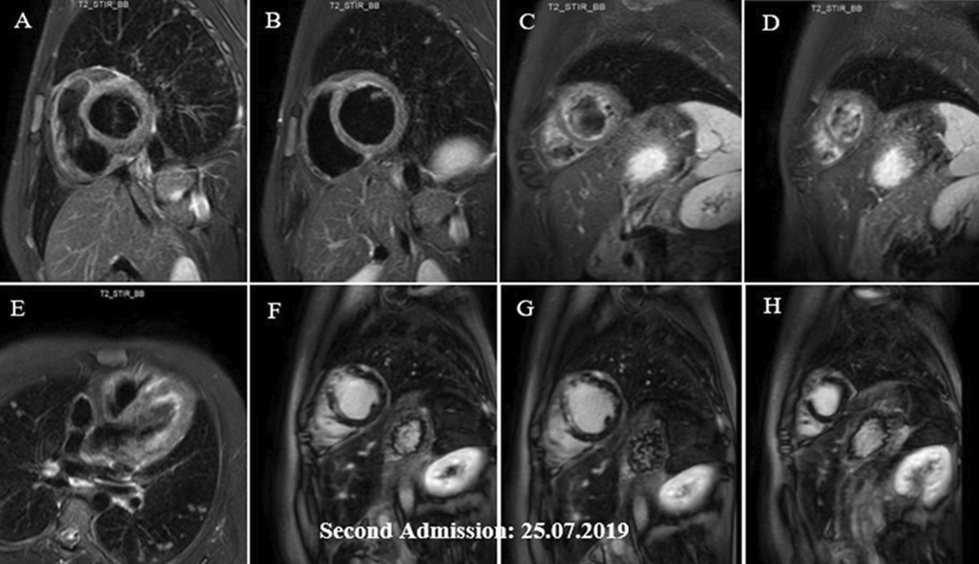
Fig. 5.

A–H Short axis basal, mid, apical and 4-chamber STIR images demonstrating subendocardial and with lesser degree midmyocardial and epicardial edema in different LV walls (mainly the apex) and also in RV walls (A–E), which has decreased compared with previous study (shown in Fig. 4A–C), indicative of partial improvement. Short axis post contrast views showing subendocardial, midmyocardial and epicardial LGE in anterior, septal and with lesser degree anterolateral LV walls, mainly consistent with residual inflammation and with lesser degree areas of midmyocardial scar formation in anterior and septal walls (F–H)
