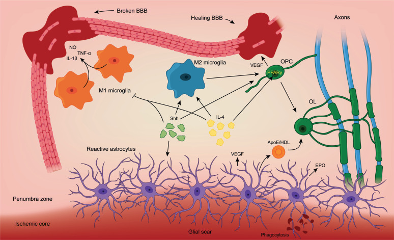
Fig. (3).
Schematic illustration of novel intercellular interactions in response to brain ischemia. Upon ischemic damage, a variety of signals is released to the medium. M1 microglia releases pro-inflammatory signals like nitric oxide (NO), tumoral necrosis factor α (TNF-α) or interleukin-1β (IL-1β) and disrupt the blood-brain barrier (BBB). These effects are mediated by NF-κΒ. M2 microglia promotes repair mechanisms and Oligodendrocyte Precursor Cell (OPC) proliferation. OPCs release vascular endothelial growth factor (VEGF) to promote angiogenesis and repair the BBB. In addition, they will differentiate into mature Oligodendrocytes (OL) to enseath damaged axons. Reactive astrocytes participate in the process by generating the glial scar to confine the ischemic core. They also provide OLs with cholesterol via Apolipoprotein E (ApoE)/ High-density lipoproteins (HDL) for myelin biogenesis and release regulatory factors like VEGF and erythropoietin (EPO). Sonic hedgehog (Shh) signals modulate glial scar formation, direct microglia towards M2 phenotype and promoting OPC proliferation. Interleukin-4 (IL-4) also guide M2 differentiation and OPC proliferation via PPARγ.

