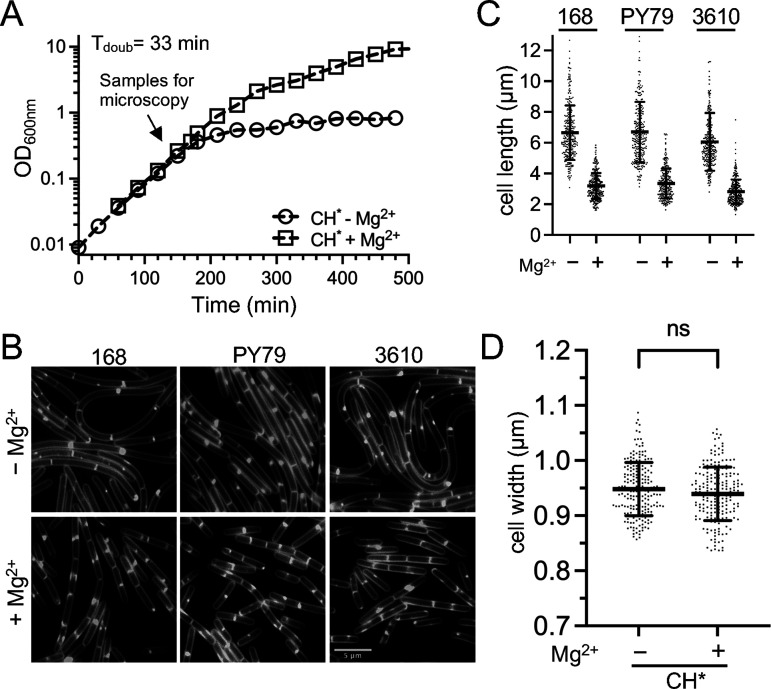
FIG 2.
Cell length of three B. subtilis strains following growth in CH* medium. WT B. subtilis 168 (BJH004), PY79 (BJH001), and 3610 (BJH403) were grown at 37°C to the mid-exponential phase in CH* supplemented with 10.0 mM MgCl2 as indicated. (A) Representative growth curves for the WT. (B) Representative micrographs following the membrane staining and epifluorescence microscopy (scaled identically). (C) Scatterplots showing the distribution of cell with quantitated for 300 cells from each condition. The bars represent the means of 300 cells ± the SDs. (D) Scatterplots showing the distribution of cell widths for 200 cells grown without or with 10.0 mM MgCl2. The bars represent the means of 200 cells ± the SDs. ns, P > 0.05.

