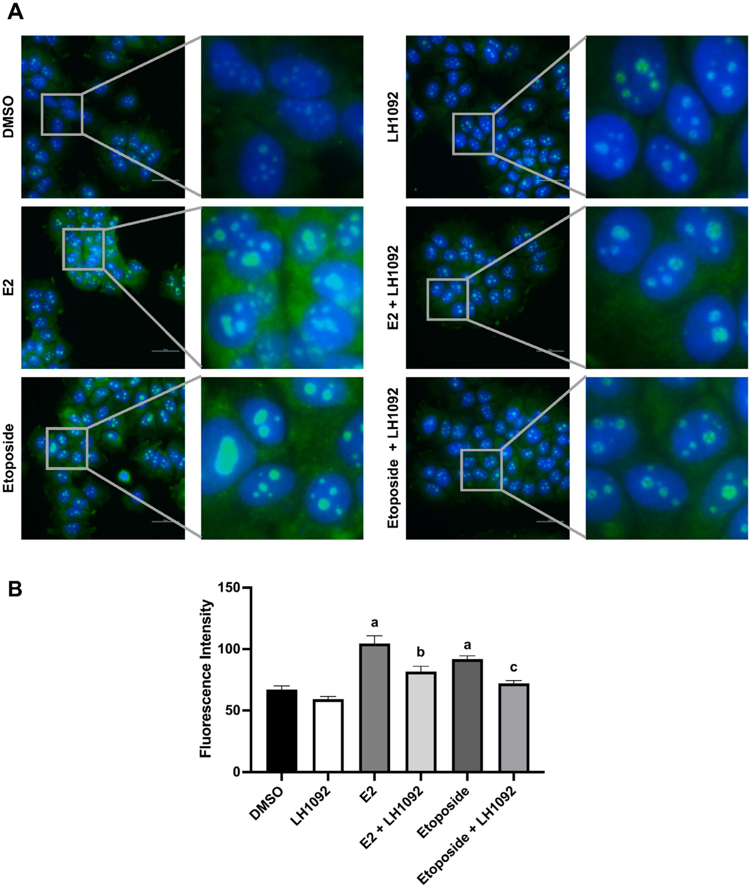
Fig. 6.

LH1092 attenuates the level of 8-oxo-dG induced by E2 and etoposide in MCF-7 cells. A. Cells were treated with 10 μM LH1092 with or without 1 nM E2 or 1 μM etoposide for 48 hours. Cells were fixed and stained for the nuclei (blue) and 8-oxo-dG (green). A fluorescence microscope was used to take the images at 40× magnification. One representative image from each group is shown here along with the expanded view. Scale bar = 50 μm. B. The immunofluorescence staining of 8-oxo-dG in the nuclei was quantified by ImageJ. Images were taken by randomly selecting five fields from each treatment group. Three independent experiments were performed. The data are represented as mean ± SEM of fluorescence intensity. a, p < 0.05, as compared to DMSO. b, p < 0.05, as compared to E2. c, p < 0.05, as compared to etoposide.
