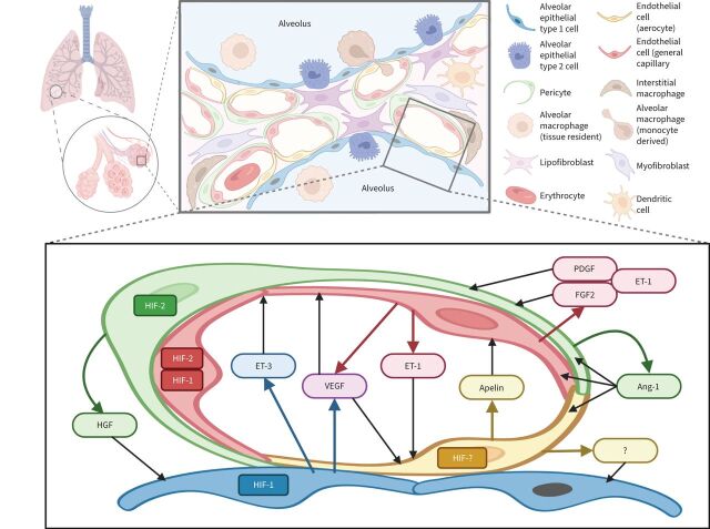
FIGURE 1.
Schematic representation of paracrine interactions within the alveolar-capillary unit of the lung acinus: focus on the microvasculature. An alveolar capillary consists of “respiratory” and “homeostatic” compartments. The first one provides gas exchange and is composed of alveolar epithelial type 1 cells, aerocytes (aCap) and a thin basal membrane between them. The second one consists of closely connected general capillary endothelial cells (gCap) and pericytes that both may contribute to structural stability, regeneration and vascular tone. Crosstalk between gCap and pericytes is mediated by angiopoietin-1 (Ang-1), endothelin (ET)-1, platelet-derived growth factor (PDGF) and fibroblast growth factor-2 (FGF2). Aerocytes are unique microvascular endothelial cells with a large surface designed for effective gas diffusion. They produce apelin that is antagonistic to ET-1 released by gCAP. In addition, a subset of gCap cells represents progenitors for both gCap and aCap. The main pro-survival and mitogenic signals for endothelial cells within the alveolar-capillary unit are vascular endothelial growth factor (VEGF) and Ang-1, produced by alveolar epithelial type 1 cells and pericytes, respectively. Secretion of most of the angiocrine factors is hypoxia-inducible factor (HIF)-dependent: HIF-2α is a predominant form in pericytes and gCAP, while HIF-1α is predominantly expressed in alveolar epithelial type 1 cells. HIF isoforms in aerocytes and factors that may affect alveolar epithelial cells have not been described. HGF: hepatocyte growth factor.

