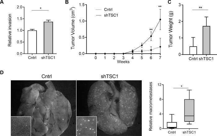
Fig 4. Independent silencing of TSC1 promotes invasion and metastasis.
(A) Relative invasion of LMS cells transduced with shTSC1 or non-target shRNA control (Cntrl) in transwell assay (n = 3). Cells were counted on membrane inserts after crystal violet staining. Cell counts were normalized to the control group. (B) Primary tumor growth following subcutaneous injection of control (n = 10) and shTSC1 (n = 10) transduced LMS1 cells in immunocompromised mice. (C) Mean weight of primary tumors at endpoint (7 weeks) for control (n = 6) and shTSC1 (n = 8) groups. (D) Fluorescent images of GFP positive nodules in whole lungs (left panel). Box plot (right) depicts average number of GFP+ lung macrometastases in 3 randomly chosen fields (inset) for Ctrl (n = 10) and shTSC1 (n = 10) xenografted tumors. Statistical analyses were performed by two-tailed students t-test; *P < 0.05, ** P < 0.005.

