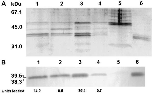
FIG. 3.
Sodium dodecyl sulfate-polyacrylamide gel electrophoresis (SDS-PAGE) and Western blot analysis of Hap production. An 8-h culture of strain C7258 was divided into aliquots, the cells were resuspended in 1 volume of the different media described below, and incubation continued at 30°C with shaking (200 rpm) for 4 h. The cultures were centrifuged, and 2 ml of each supernatant was concentrated 10-fold by centrifuging it through Centricon-10 centrifugal filters. The retentate was adjusted to 0.2 ml, and 0.02-ml aliquots were analyzed in an SDS–12% PAGE gel. (A) SDS-PAGE. Lane 1, cells resuspended in the original 8-h spent medium; lane 2, cells resuspended in 1× fresh TBS; lane 3, cells resuspended in 0.25× fresh TBS; lane 4, cells resuspended in 0.25× fresh TBS supplemented with 0.4% d-glucose; lane 5, concentrated supernatant of mutant 638; lane 6, pure Hap (2.5 μg). (B) Western blot. Identical samples were loaded in a second gel and transferred to a polyvinylidene difluoride membrane. Hap was detected by using a rabbit anti-Hap serum and peroxidase-conjugated goat anti-rabbit immunoglobulin G. The total azocasein units loaded per lane are indicated below the blot. Molecular masses were calculated with reference to SDS molecular mass standards (low range) from Bio-Rad laboratories.

