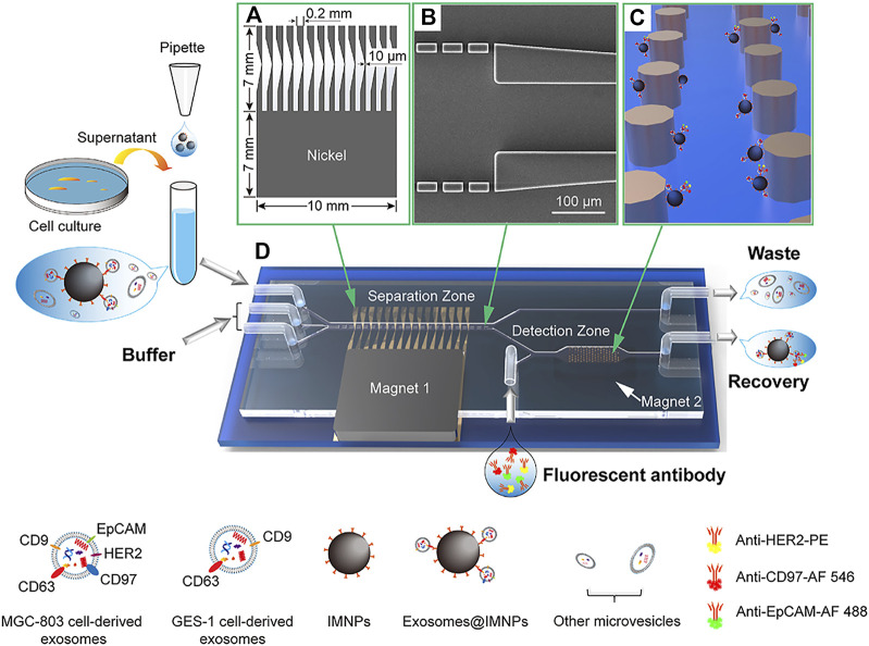
FIGURE 7.
ExoSD chip for exosome separation by IMNPs. The separated exosomes@IMNPs were captured on the Ni cylinder array and labeled with fluorescent antibodies for detection. (A) Scheme of the Ni comb-like structure for local magnetic field and magnetic field gradient enhancement. (B) SEM image of the microfilter. (C) Schematic diagram of capturing exosomes@IMNPs by Ni cylindrical array. (D) Three-dimensional diagram of the ExoSD chip. Reproduced with permission from (Yu et al., 2021).

